An operation attracting significant international interest was performed on May 6, 2026, at Semmelweis University’s Department of Obstetrics and Gynecology, an event that also marked a new phase in the collaboration between the organizational unit and the Department of Neurosurgery and Neurointervention. A rare, benign tumor originating from the sheath of peripheral nerves, known as a schwannoma, was removed from a young woman’s lesser pelvis by the department’s neuropelveology team, in close collaboration with the neurosurgical and intraoperative neurophysiology working group led by Dr. Loránd Erőss, Director of the Department of Neurosurgery and Neurointervention, along with electrophysiologist Dr. Boglárka Hajnal and neurology resident Dr. Borbála Damó-Csorba.
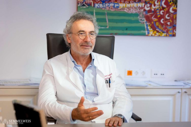 “In this case, we encountered a very rare variant of schwannoma: A tumor growing in the retroperitoneal space behind the uterus in the lesser pelvis, which accounts for 1–3 percent of all schwannomas,” Dr. Loránd Erőss told our website. As he pointed out, such cases require particularly thorough neurological or spinal surgical evaluation, as the severe pain that occurs in these instances – in the sciatic nerve and the sacrum, and radiating to the lower extremities – is most characteristic of degenerative spinal diseases, which are considered a widespread condition, so the cause is usually sought there.
What is a neurinoma or schwannoma?
A neurinoma, a slow-growing tumor that develops from the myelin sheath of nerves, is a rare condition, with an incidence of 0.3–0.5 cases per 100,000 people; however, it can develop anywhere in the body where there are peripheral nerves: in the limbs, nerve plexuses, nerve roots exiting the spine, the auditory nerve, and other cranial nerves. The most common is the vestibular schwannoma, a benign tumor of the Schwann cells of the eighth cranial nerve, accounting for 6–10 percent of all intracranial tumors.
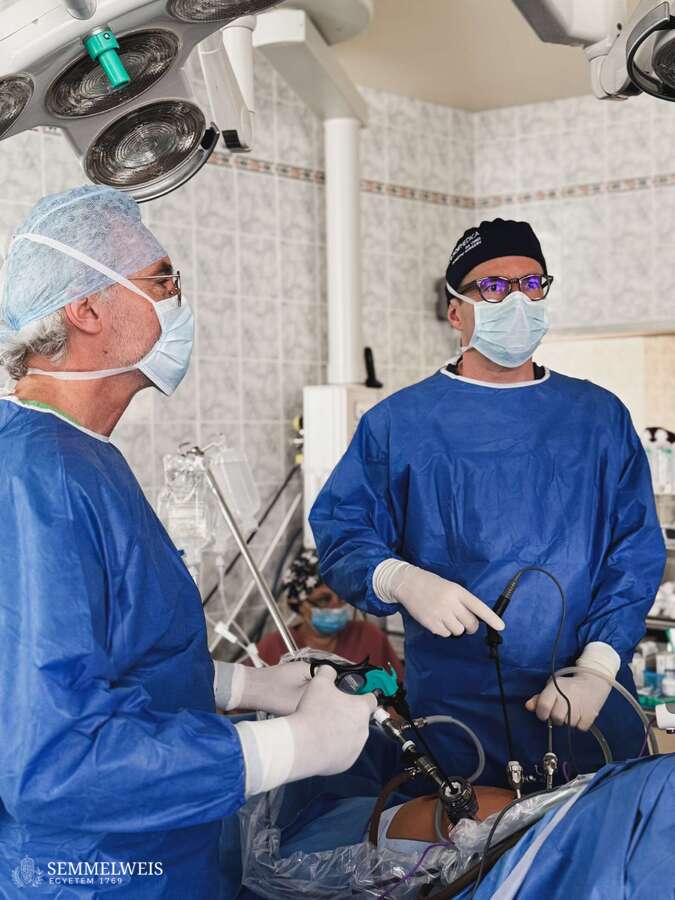
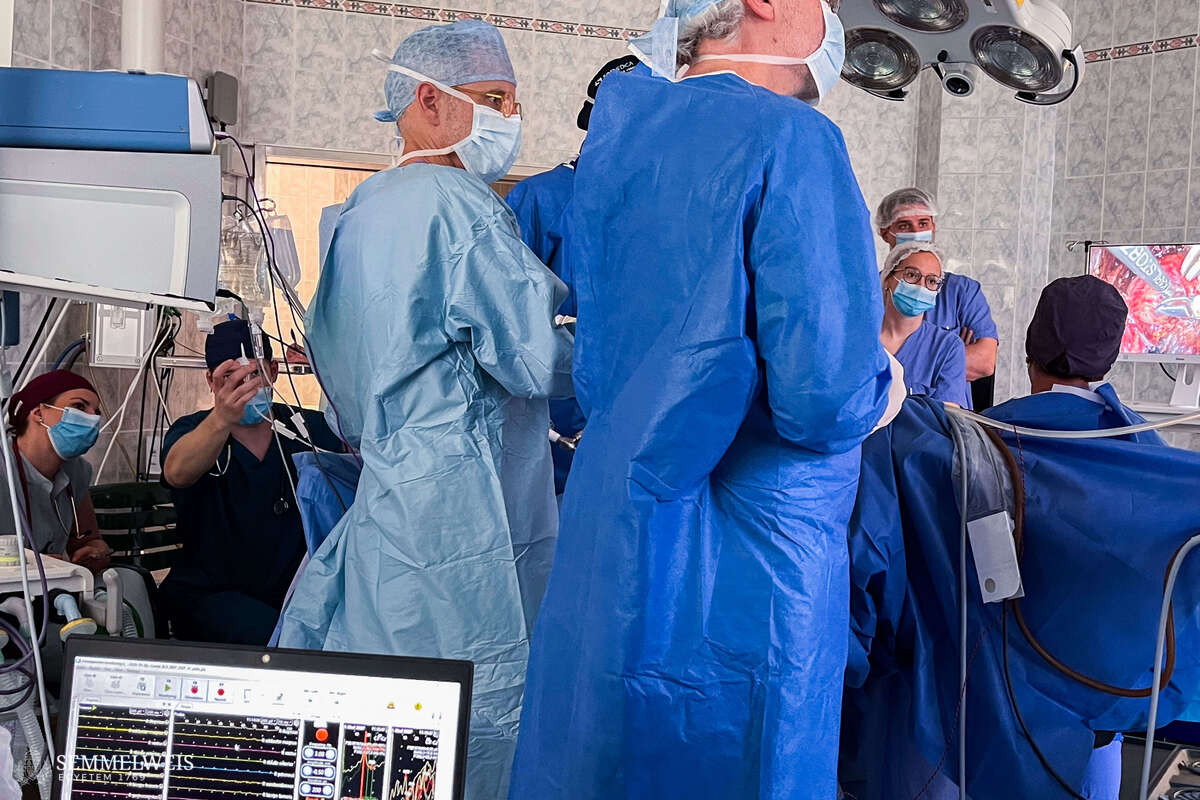
“In this patient, a five-centimeter tumor developed behind the uterus at the point where the nerve exits the sacrum, compressing the nerve; the patient sought examination due to unrelenting pain radiating into the right leg,” said Associate Professor Dr. Attila Bokor, Head of the Endometriosis Center and the laparoscopic surgery unit at the Department of Obstetrics and Gynecology. Recognizing that this was a unique clinical picture, and that extreme caution was required during the procedure due to the location in the lesser pelvis and the need to preserve the function of the sensitive nerves and organs in that area, he asked the surgeons at the Department of Neurosurgery and Neurointervention to collaborate and to provide electrophysiological monitoring during the operation.
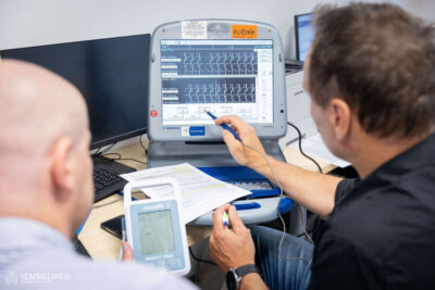
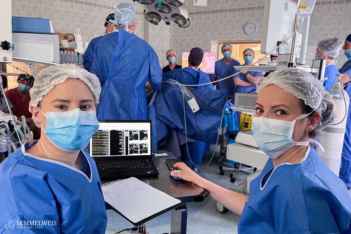
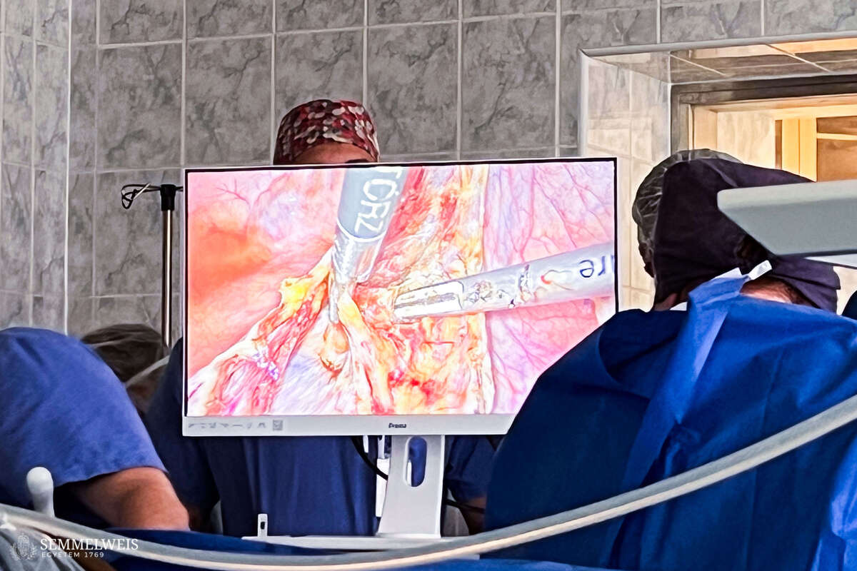
 As Dr. Attila Bokor explained, during the approximately five-hour surgery performed at the Baross Street section of the Department of Obstetrics and Gynecology, the neurosurgeons continuously monitored the surgical site using a 32-channel electrode and a connected neuromonitor to determine whether the area being operated on contained any nerve bundles, or only the wall of the neurinoma, and whether removing that tissue would cause nerve damage or not. “From a neurosurgical perspective, the challenge of the surgery was the laparoscopic localization of the tumor, but Dr. Attila Bokor’s laparoscopic experience managed to overcome it,” said Dr. Loránd Erőss. Using neurostimulation performed during laparoscopic dissection, they precisely localized the nerves running along the surface of the tumor; thus, after dissecting the nerve bundles running along the surface of the tumor, they were able to remove it in several pieces.
This provided a great deal of safety during the surgery, as a tumor approximately five centimeters in size had to be removed from an area that was particularly difficult to access and prone to bleeding. – Dr. Attila Bokor
An additional challenge of the procedure was that the tumor was not only compressing the nerve of origin, but it had also begun to affect the function of another nerve root. Moreover, the nerves controlling the function of the rectum, bladder, and vagina also run through this area. Preserving these functions, as well as uterine fertility, and avoiding neurological deficits, were among the goals of the surgery, in addition to eliminating pain and removing the tumor, as Dr. Attila Bokor noted.
“To facilitate long-term collaboration and ensure the successful execution of joint surgeries, we have also acquired a mobile neuromonitoring device,” added Dr. Loránd Erőss. This is used, among other things, when removing endometriotic lesions that affect nerves in the lesser pelvis, as a delicate balance must be struck between excising the lesions causing pain, thus sacrificing some of the nerves that regulate various vital functions, and preserving those functions. The device will also be useful in treating various vascular compressions in the lesser pelvis that develop as a result of previous surgeries, radiation therapy, or conditions associated with significant fibrosis and inflammation.
“These are rare conditions; we perform 2–3 such planned surgeries every three months, which makes us the only ones in Hungary and neighboring countries to do so. Even on a European scale, only one French and one Swiss working group perform a similar number of such neuropelveological procedures,” added Dr. Attila Bokor. For this very reason, the neurinoma removal performed in early May with intraoperative electrophysiological monitoring attracted interest not only from within the university; the operation was also watched live by specialists from Slovenia, Turkey, and Portugal, among others.
Gallery
The surgery went according to plan, and the patient was discharged from the department the next day. Not only was she relieved of her pain, but also of the risk of lower limb paralysis. “She has now fully recovered; going forward, she only needs to undergo follow-up MRI scans at three months and one year,” added Dr. Loránd Erőss.
Melinda Katalin Kiss
Translation: Dr. Balázs Csizmadia
Photos by Bálint Barta, Boglárka Zellei, Dr. Attila Bokor, Dr. Boglárka Hajnal – Semmelweis University