Among the diseases affecting the aorta, the body’s main artery, the most common is aneurysm, or the dilation of a blood vessel, which most often develops in the abdominal section. If the dilation reaches a certain size – on average, a diameter of 5.5 centimeters – surgery is the only way to prevent a life-threatening condition from developing. The intervention can be either open surgery or a minimally invasive endovascular procedure, which is less demanding for the patient. During the procedure, a stent graft is inserted into the aneurysmal segment of the artery via the femoral artery, which takes the strain off the dilated vessel. From a surgical standpoint, this is a complex procedure in which the surgeon does not have a direct view of the surgical site but relies solely on the imaging device. In addition, the shape and location of the aneurysms can vary greatly.
This is precisely why various surgical planning software programs are so useful, as they help to simulate the surgery in advance, tailored to the patient’s specific anatomy, and to determine the exact size, placement, and type of the stent graft to be implanted.
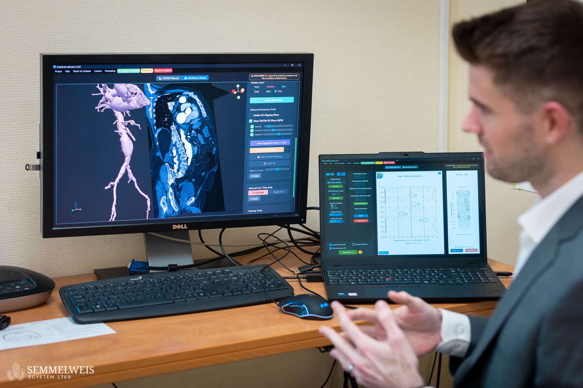
One such piece of software, currently available for educational and research purposes, was developed by Bendegúz Juhos, a certified electrical engineer and PhD student at the Department of Interventional Radiology of the Városmajor Heart and Vascular Center, under the supervision of Dr. Csaba Csobay-Novák.
 “I joined the program as an engineer – a unique path – and became a member of the group focusing on aortic diseases. “As part of one of my research projects, I developed the EndoDraft surgical planning software, originally designed specifically for complex abdominal aortic aneurysm stent graft procedures, which is extremely effective in assisting specialists with planning and with applying the stent graft modification techniques developed at our department,” explained Bendegúz Juhos.
What is a stent graft?
A stent graft is a biocompatible, flexible, tube-like material reinforced by a metallic mesh (strut). It is used to treat diseases of blood vessels (most commonly the aorta). It is inserted into the blood vessel via a catheter, where it is secured to the vessel wall at the appropriate location and relieves the pressure on the dilated vessel.
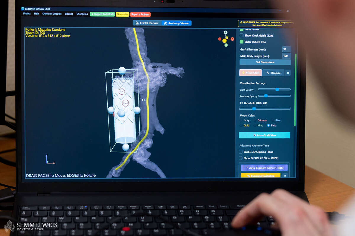
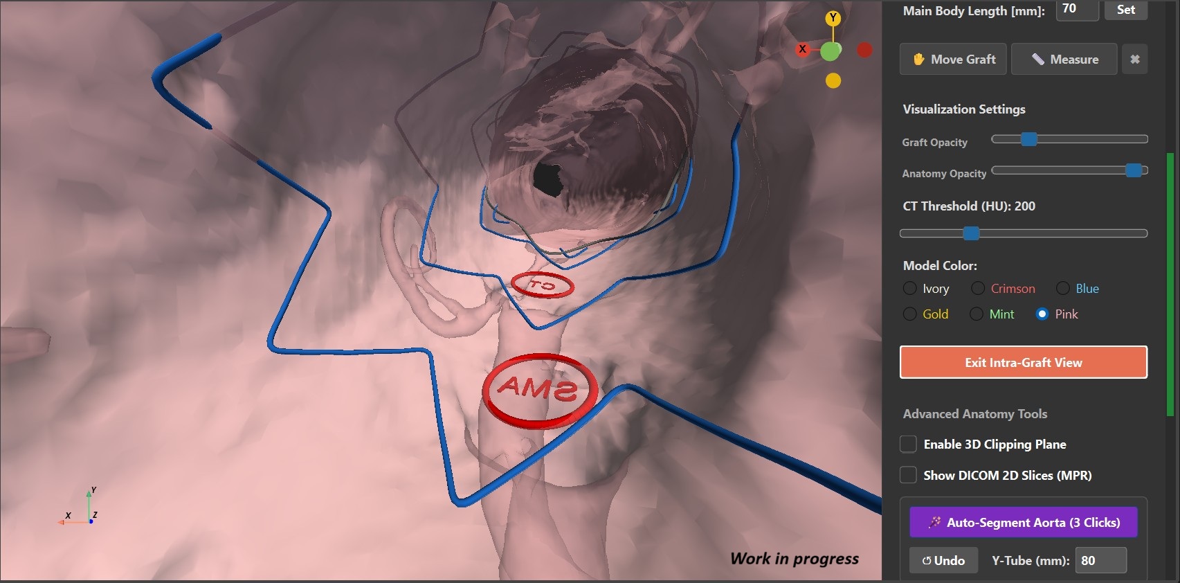
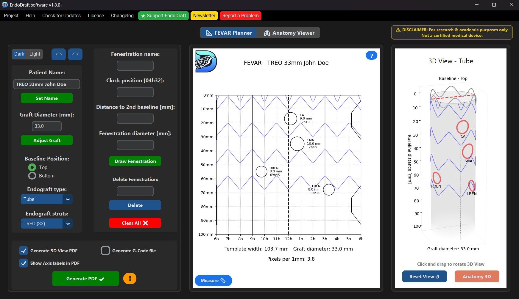
“In emergency or acute surgeries, the physician may need to modify the commercially available stent graft due to the patient’s unique anatomical characteristics (Physician-Modified Endograft; PMEG). Surgical planning software – including the clinic’s own software – can assist with this: It calculates with millimeter accuracy where holes need to be made in the graft, and a precision printing template (laser foil) can also be created, which helps ensure the accurate creation of these so-called fenestrations; furthermore, the software allows for the export of patient-specific spatial anatomical models for 3D printing,” added Bendegúz Juhos. The software is not only available free of charge but also has the unique feature that the precise structure of stent grafts commonly used by various manufacturers (such as the reinforcing metallic mesh) is visible even during the planning phase. This means that it can be determined in advance, before surgery, whether a planned opening would collide with the metal frame of the device.
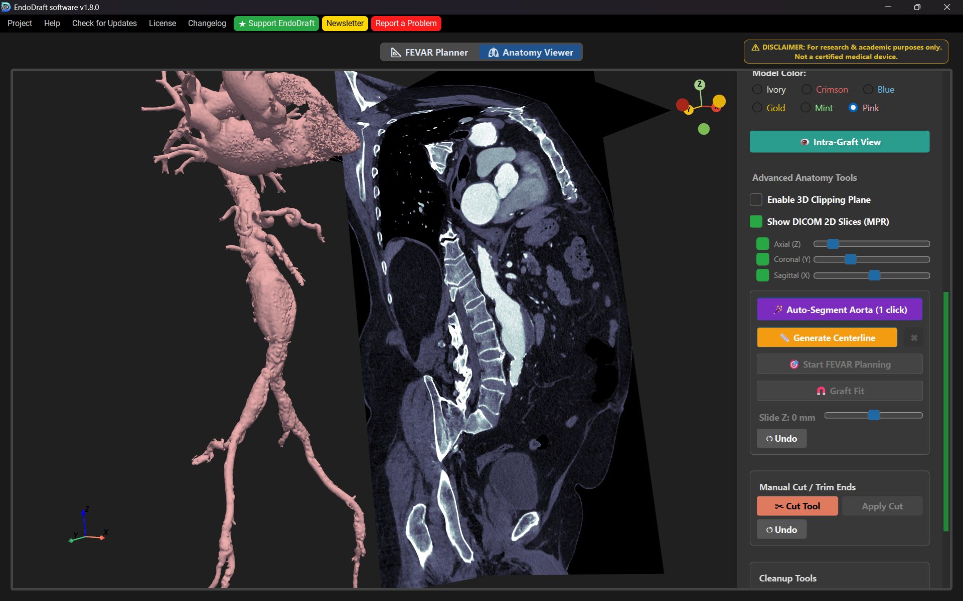
The software was recognized as an intellectual creation by the University Innovation Committee, and the name EndoDraft was trademarked; furthermore, an article on the subject was published in the Journal of Endovascular Therapy, detailing the related retrospective clinical trial. The software is currently available for free download. In addition to the previous, simple 2D simulation module, a complex module has now been developed that enables full 3D spatial planning; the patient’s CT scan can be uploaded to this module, and based on that, any automatic simulation, vascular enhancement, and spatial modification can be performed. “The latest module of the EndoDraft software can now be used for any endovascular procedure, making features widely available that were previously only accessible through costly, specialized software licenses. All of this is now available free of charge to anyone, both domestically and internationally, for educational and research purposes,” emphasized Bendegúz Juhos, who is confident that the surgical planning software will become increasingly widely used among interventional radiologists and vascular surgeons both within the university and at the national and international levels.
Gallery
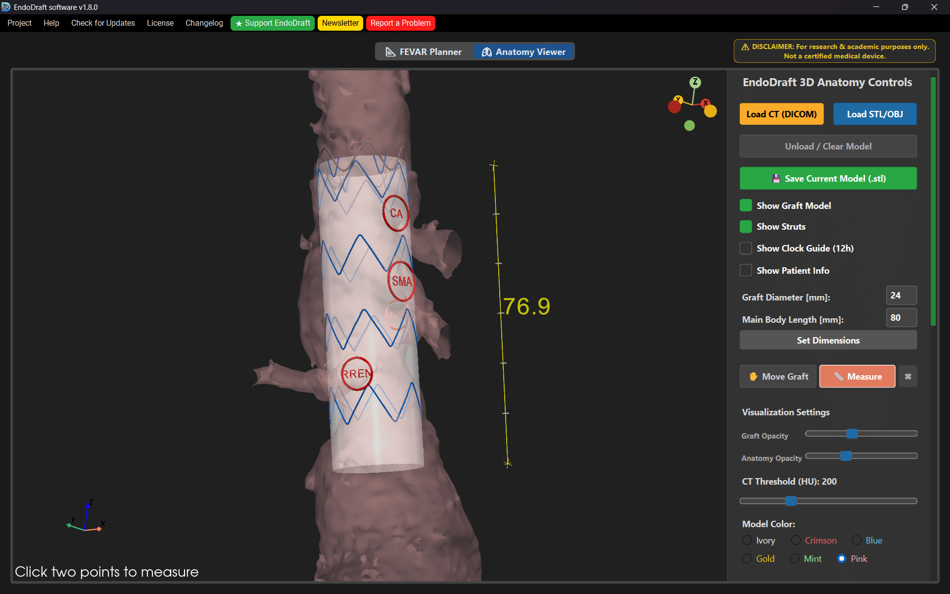
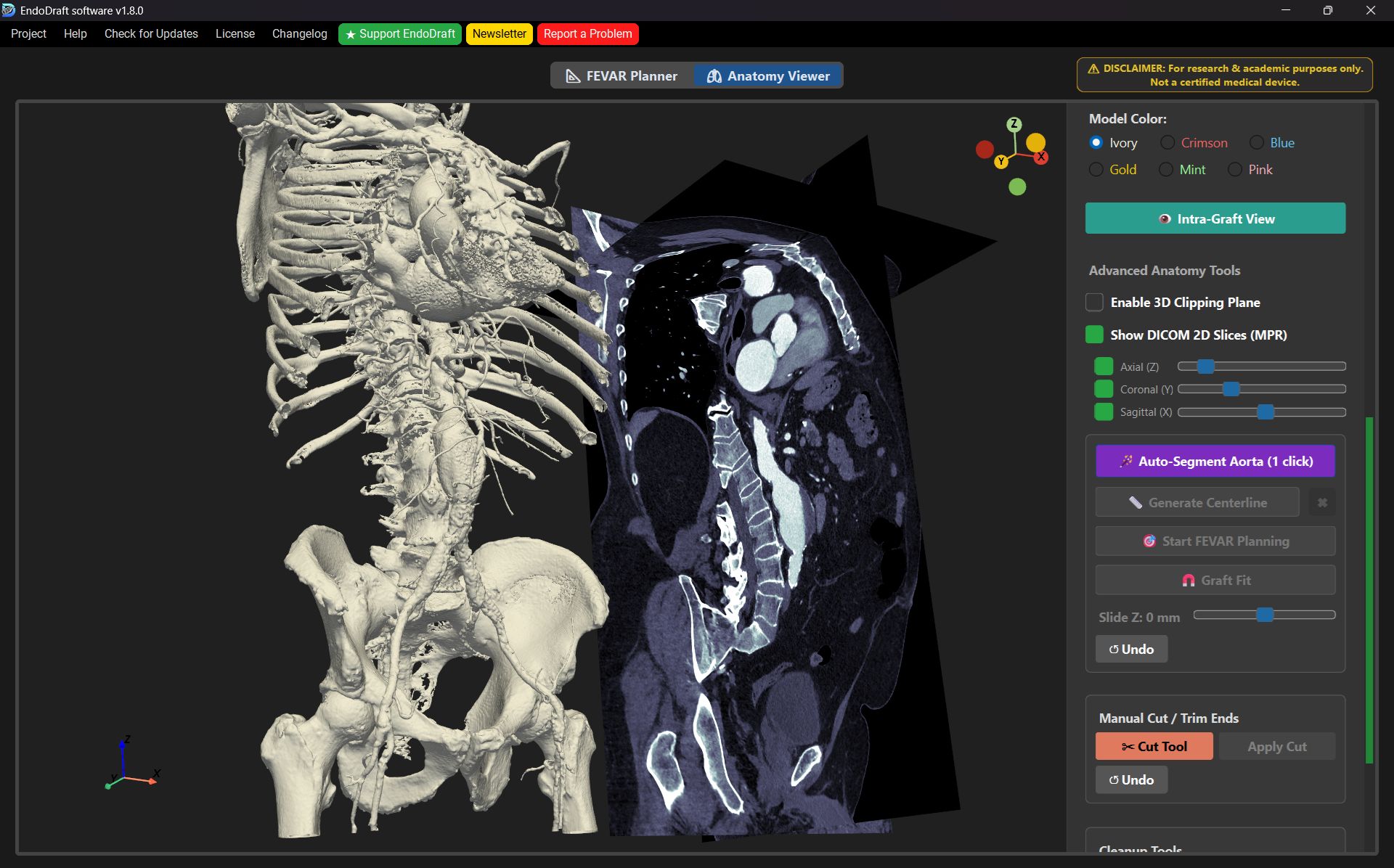
As an example, he explained that the device could be used for demonstration purposes during training; by loading a given patient’s CT scan, the instructor could show students a 3D model of what a stent graft placement looked like and how the device would be positioned and fitted.
Pálma Dobozi
Translation: Judit Dőtsch
Photos by Boglárka Zellei – Semmelweis University; illustrations: Bendegúz Juhos – EndoDraft