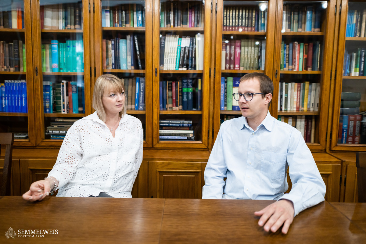
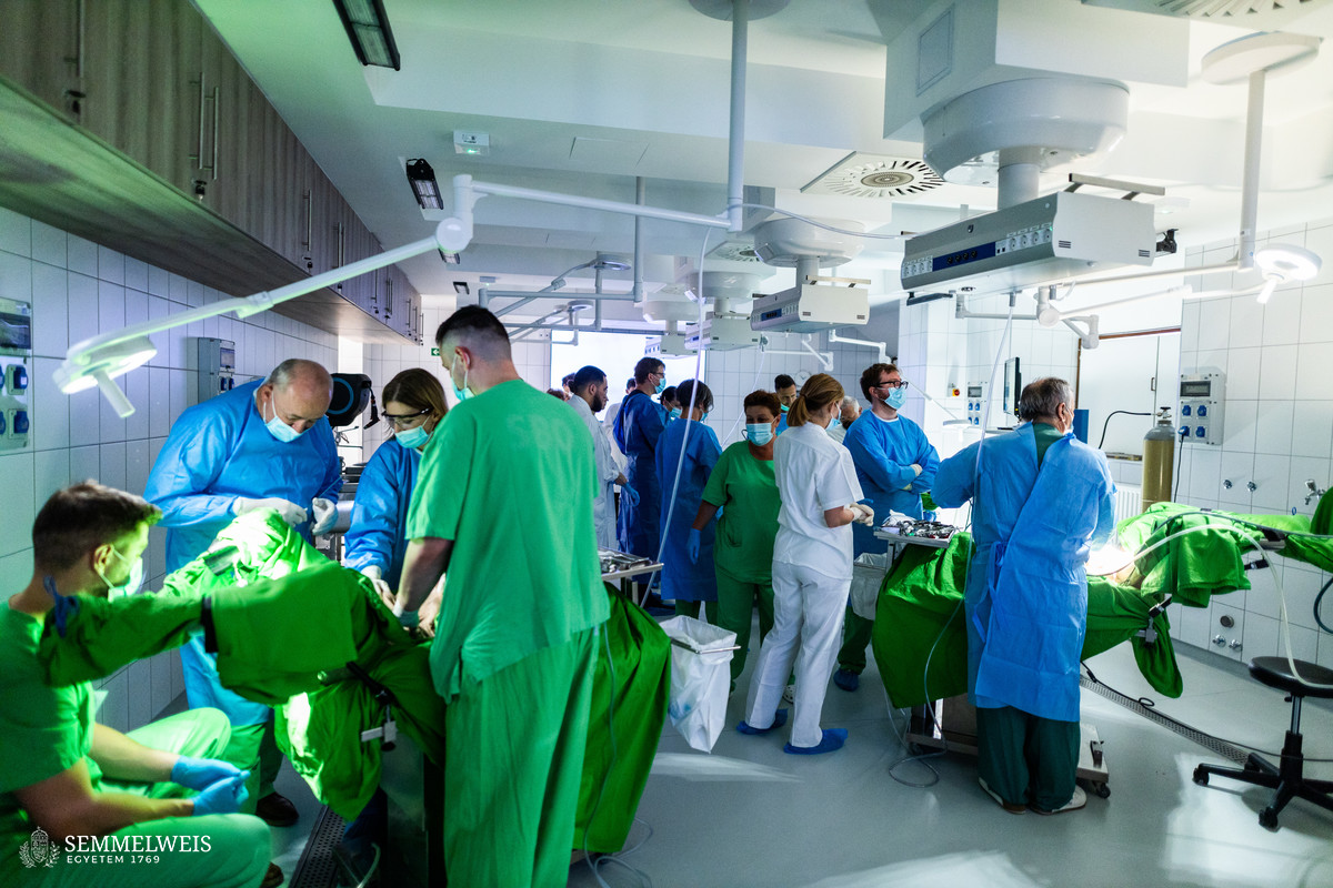
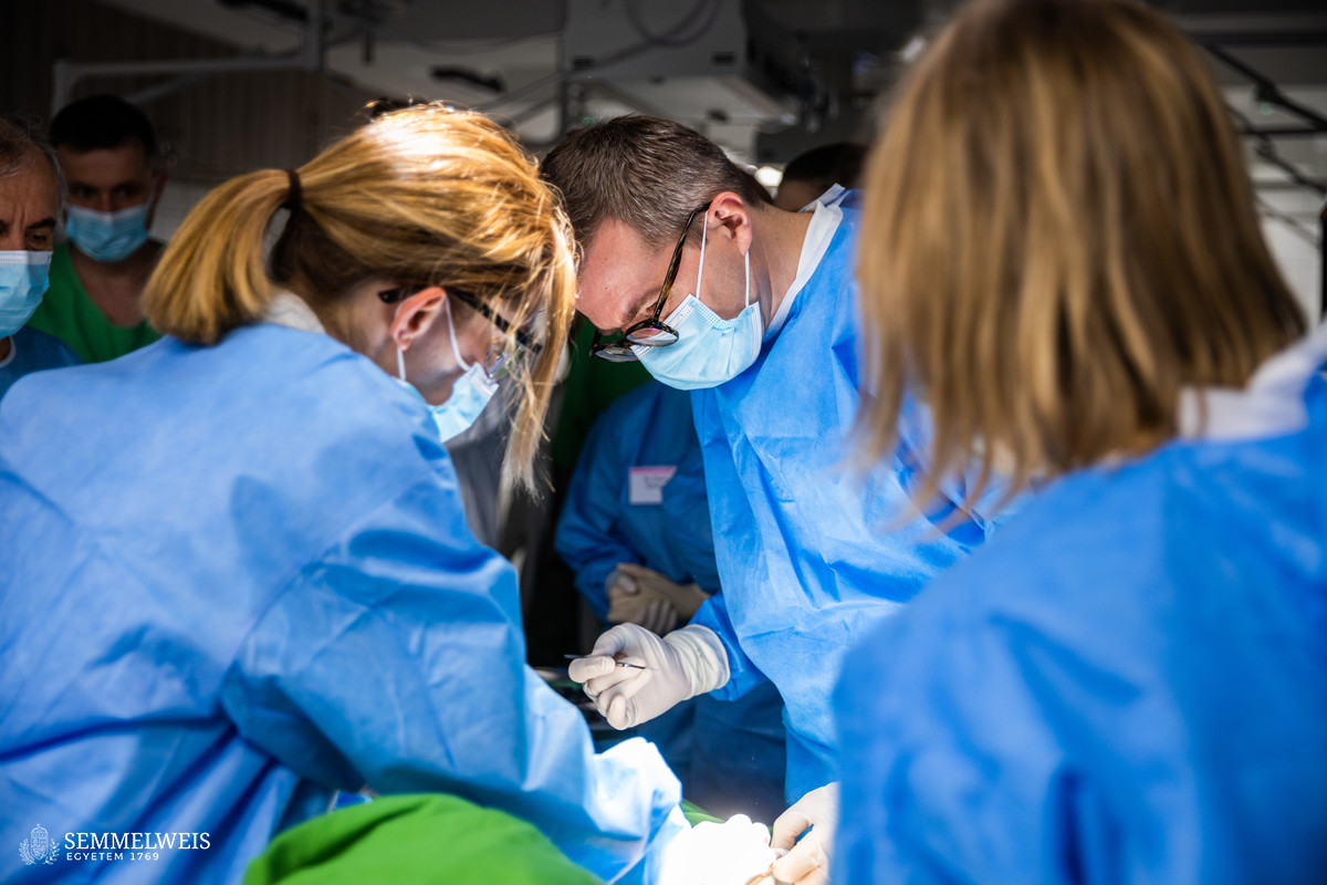
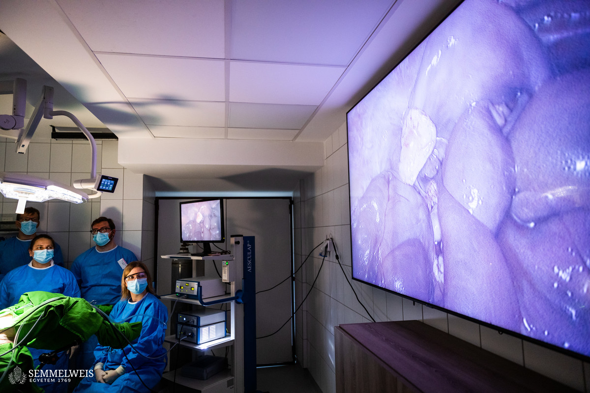
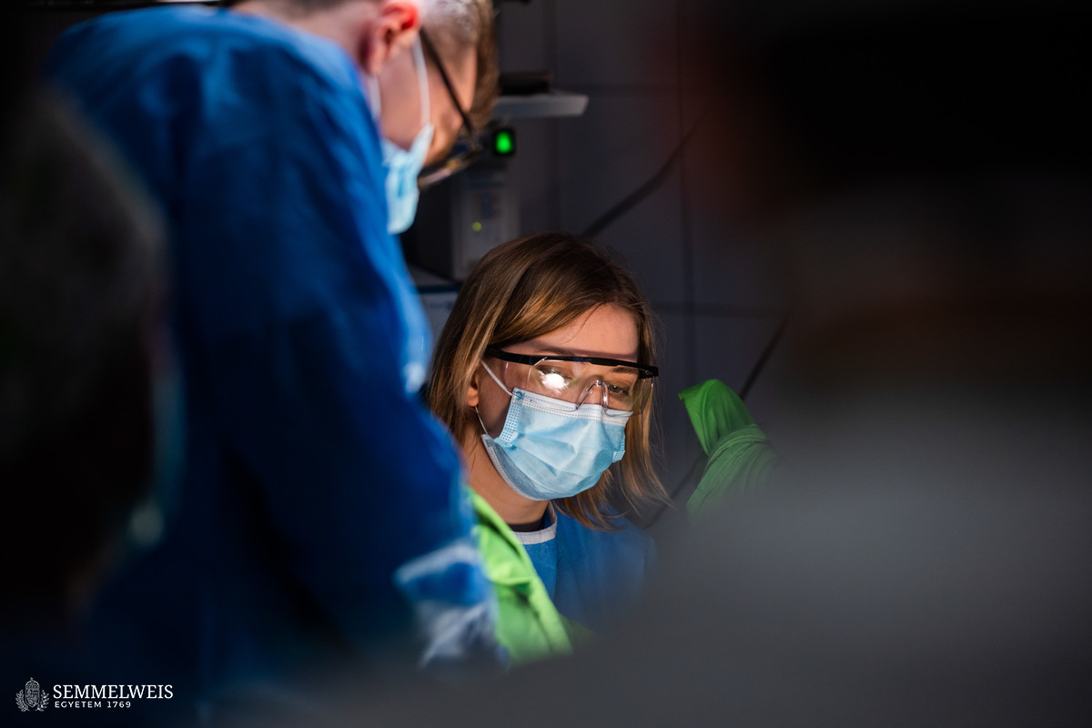
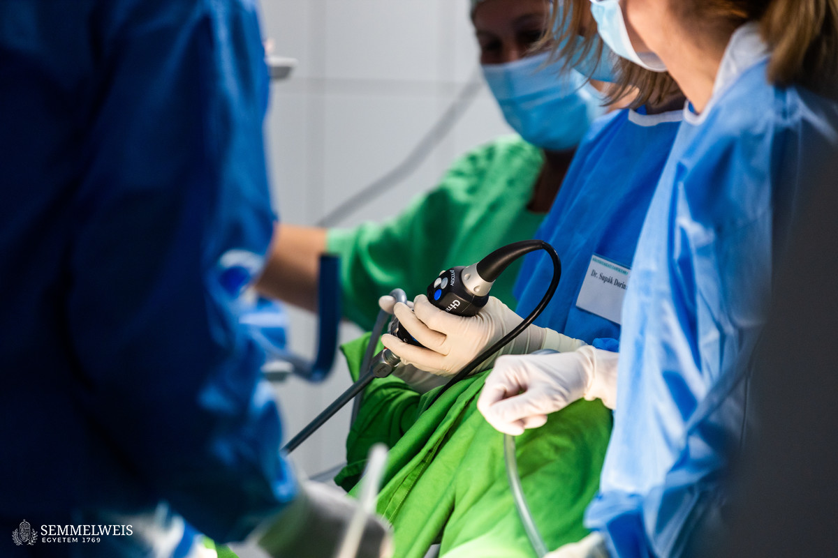
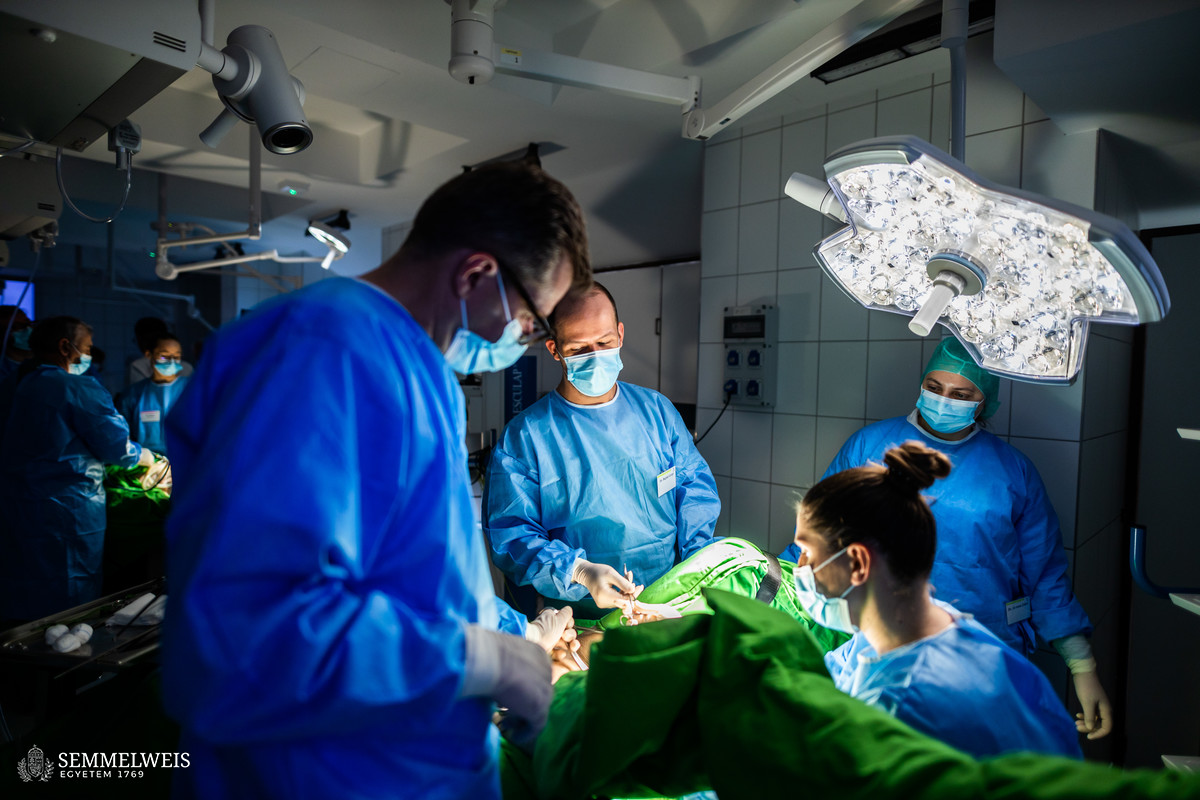
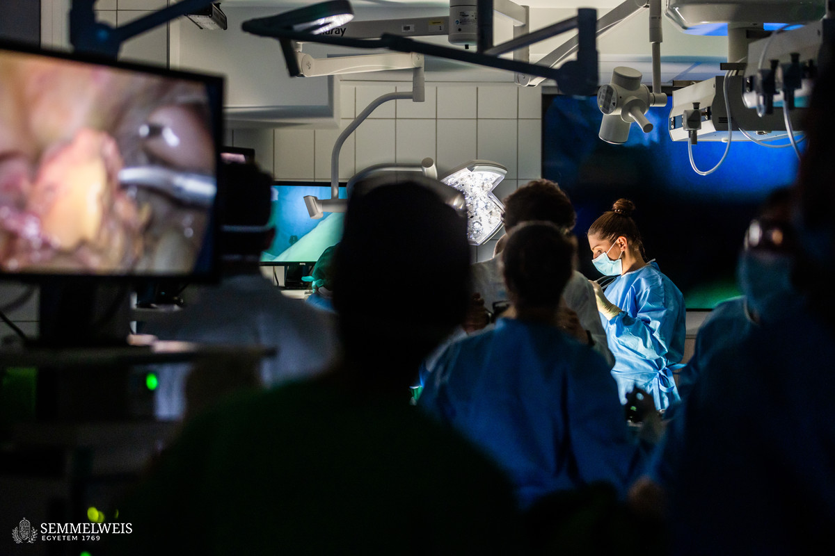
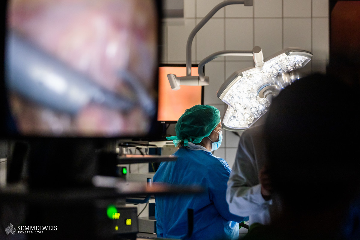
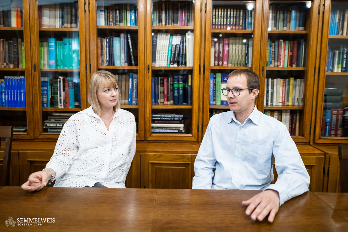
The most modern way to perform hysterectomies for benign diseases is the laparoscopic option, as this procedure is more beneficial for patients in many aspects. The only way to learn the manual skills needed to recognize and avoid the rare but very severe complication of hysterectomy, namely ureteral injury, has been to practice on surgical female pelvic trainers, animal models and assisting in laparoscopic surgery. The practice of cadavers (human corpses) providing a realistic tissue environment had so far been possible in foreign courses only, up until the Interdisciplinary Cadaveric Operating Room, inaugurated on 24 November last year, created the necessary infrastructure, told Dr. Noémi Csibi, Assistant Lecturer at the Department of Obstetrics and Gynecology, the initiator and developer of the laparoscopic gynecological surgery course. “Together with the expertise accumulated at the Department of Anatomy, Histology and Embryology, the cadavers donated for teaching and research purposes pursuant to the Health Act and the Senate’s regulations on the treatment of cadavers have created a huge opportunity for postgraduate education and for the organization of the much needed cadaveric surgery courses,” added Dr. Tamás Ruttkay, Senior Lecturer at the Department of Anatomy, Histology and Embryology.
The entry requirement for this landmark course was a basic level of proficiency in laparoscopic surgery, with a history of at least 25 such surgeries assisted or performed in order to get the most out of the course, explained Dr. Noémi Csibi. She emphasized that orientation in the lesser pelvis and the manual skills required for laparoscopic surgery can and should be acquired through many hours of practice with surgical trainers, video courses, animal surgery or assisting. These pave the way for cadaveric surgery, which is the highest level of training, an exceptional training option with its real human anatomy and tissue environment.
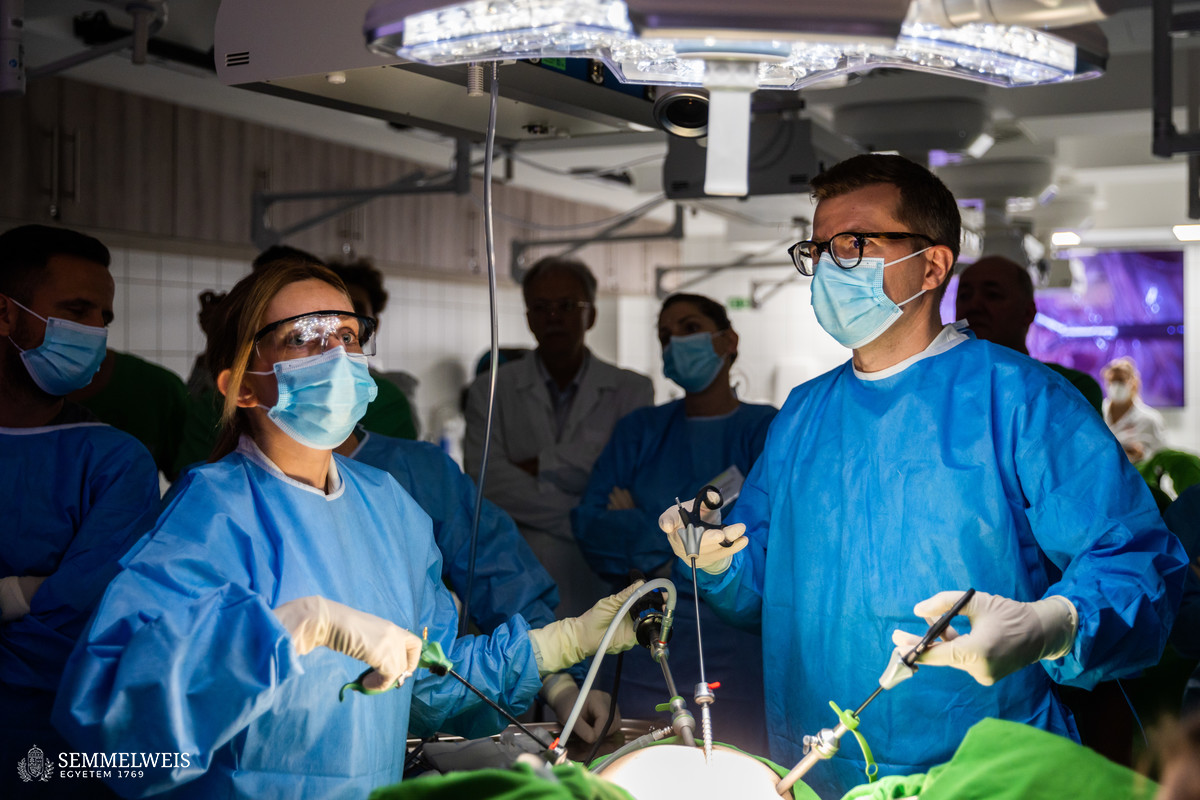
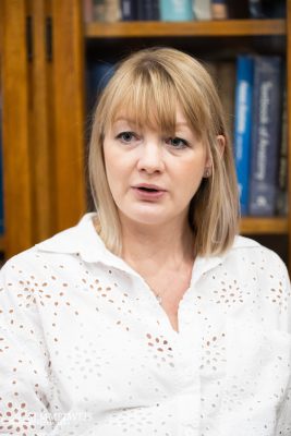 This course was not aimed at practicing the basics of laparoscopic hysterectomy, but rather at preventing a not very common but all the more dreaded complication, ureteral injury. “Our goal was to enable our students to accurately assess and become proficient in identifying when it is appropriate to access and clear the ureter, as this is rarely performed routinely,” explained Dr. Noémi Csibi. “Repairing the ureter requires wide experience and may be necessary in complicated surgical situations, so the course tried to focus in part on how a novice surgeon can recognize upcoming complications. With proper training, the right preparation, assistance and equipment, the outcome of the operation will be much more favorable than if being caught by surprise. We tried to draw attention to this in the 3-hour theoretical part of the course preceding the practice, but the main aim was to learn and practice the safe performance of an intermediate-level surgery. As far as I know, no such course in gynecological cadaveric surgery has ever been held in Hungary,” Dr. Noémi Csibi noted.
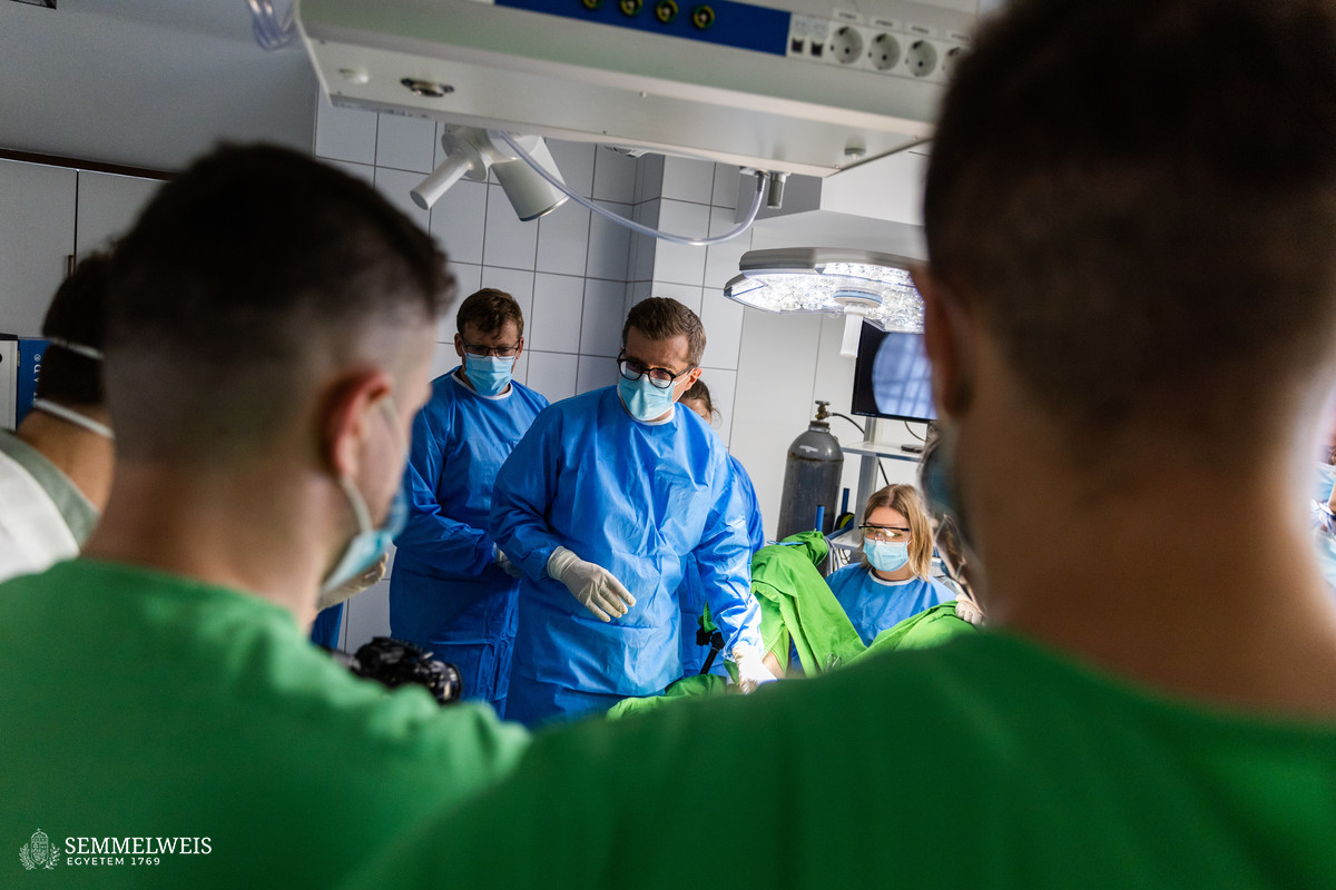
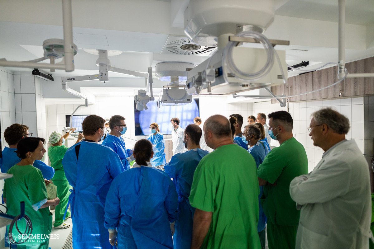
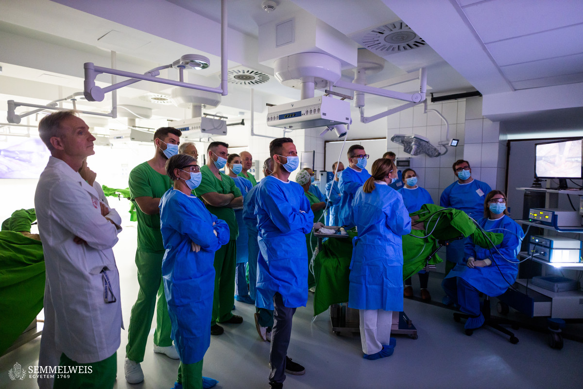
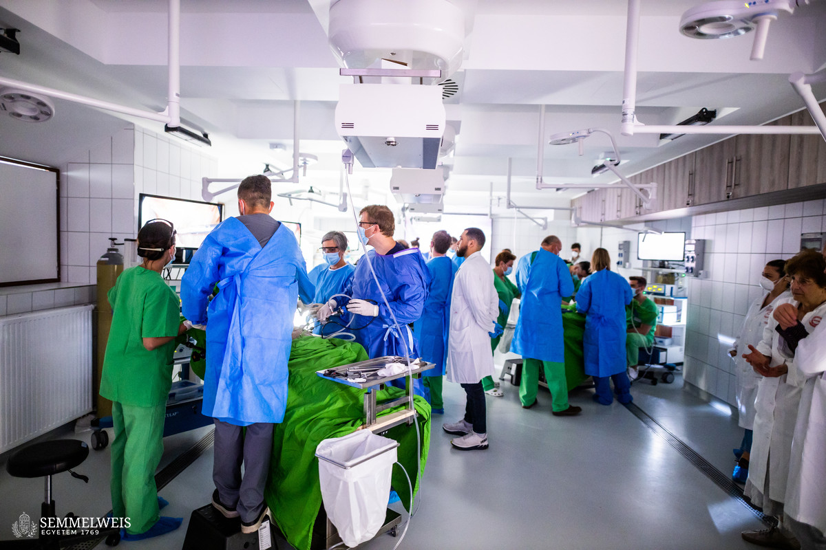
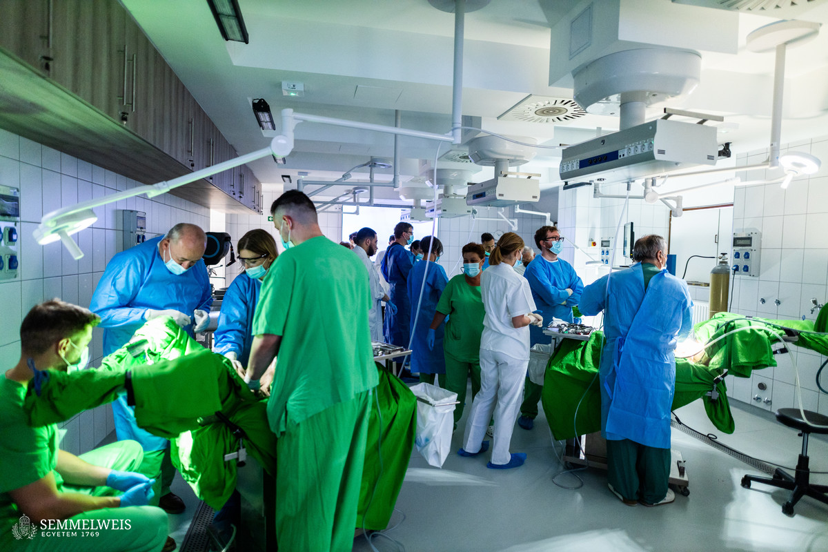
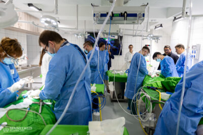
“The number of workstations was optimized jointly, so in the end the twelve participants, of whom seven were university employees and two thirds were specialist candidates, could practice on four workstations with the assistance of one instructor each, elaborated Dr. Tamás Ruttkay on the technical conditions. “By the end of the training day, feedback showed that everyone had satisfied their anatomical and practical knowledge needs to the fullest and passed the exam closing the accredited course,” said Dr. Noémi Csibi, adding that it is already clear how high the demand for such a training is.
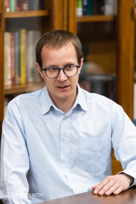 However useful cadaveric surgical training this may be, similar courses are not easily organized worldwide due to high unit costs, low number of cadaver donations and regulations on autopsy and corpse preparation, Dr. Tamás Ruttkay stressed. On the other hand, the favorable domestic legislative environment and the new interdisciplinary operating room offer bright prospects for the training of physicians in Hungary, which we plan to make unique worldwide by using disease models. This is an added bonus that is practically unmatched anywhere else in the world as far as certain fields are concerned,” Dr. Tamás Ruttkay added.
“Our concept is to bring the cadavers into conditions that are highly similar to those of living patients facing the treatment method being practiced during the course, so that the focus can be on targeted therapeutic intervention for the disease. For example, standardized artificial tumors are placed in the desired position before the training session and then shown to the participants via radiological images, who then prepare the surgical plan with the teaching expert and can step to the same cadaver to perform the exact surgery. We may even use an external pump to set the heart in motion as if it was beating. Our aim is to provide our medical students with unique training models for their bedside work, so they can heal more effectively. None of this would be possible without the selfless donation of our fellow human beings who wished to dedicate their bodies after death to medical school education and research. We are fulfilling their request, and thus our moral duty, by training the doctors of the present and the future with an ever-expanding range of opportunities,” concluded the clinical anatomist.
Dr. Tamás Ruttkay emphasized that this process is a real teamwork, as it requires the cooperation of clinical experts, clinical anatomists, preparators, autopsists, the university and the companies manufacturing, distributing and developing the instruments. The instruments are just as important contributors to the process as the equipment the participants will encounter in the operating room since the entire project makes only sense if the students can work with the same tools, he pointed out, noting that negotiations with the companies have already started. The laparoscopy towers used during the first gynecology course were provided by the Herceghalom vivisection operating room of the Heart and Vascular Clinic. As regards further plans, seven cadaver courses on other topics have already been announced for the autumn period, and a gynecology course similar to the one in early June is for about a year’s time.
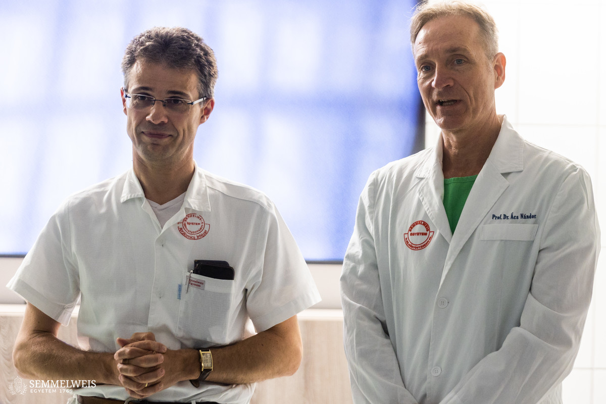
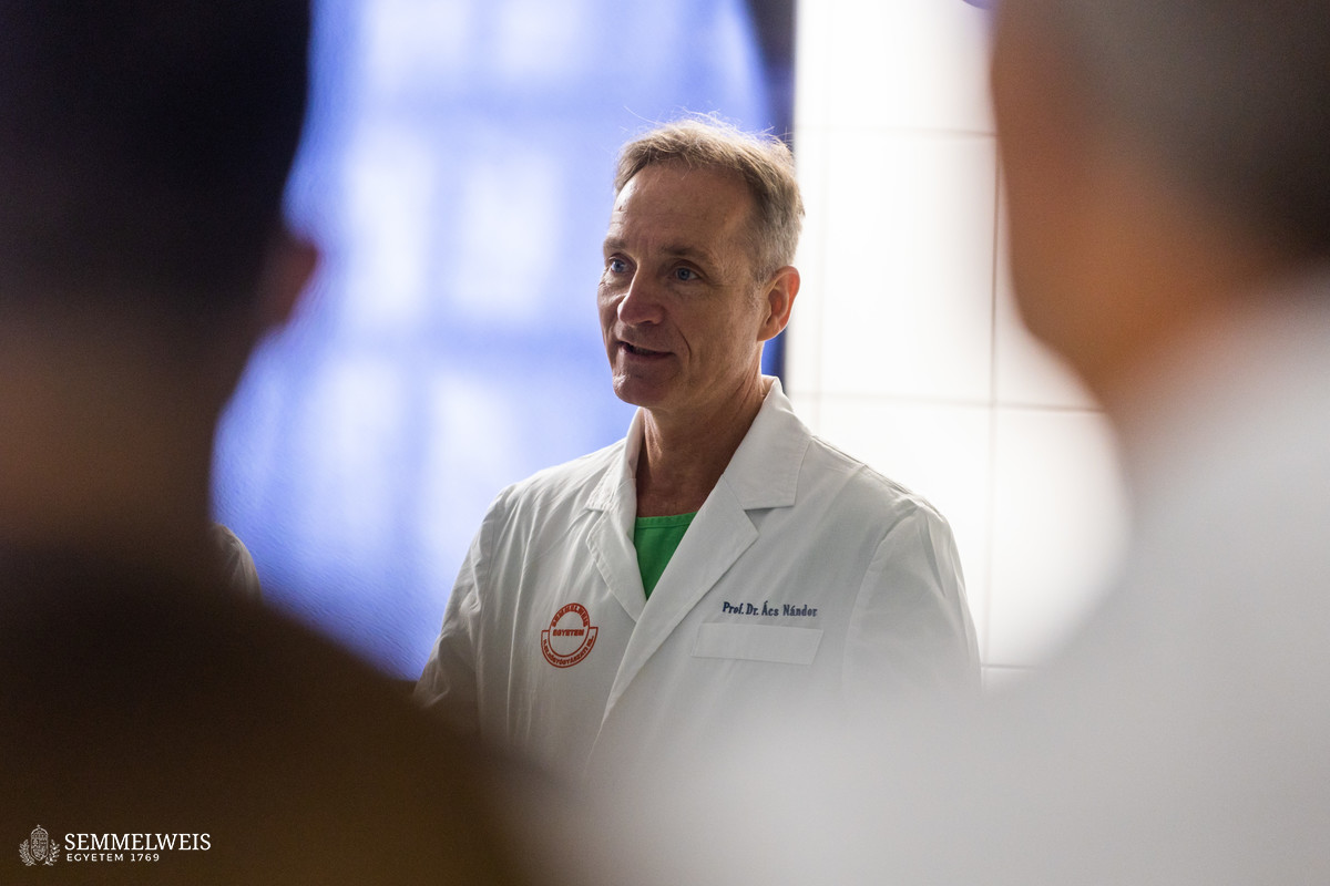
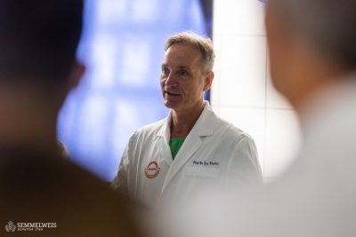 “When introducing any new surgical technique, it is best for surgeons to practice on medical simulators or animal models rather than in a real-life situation, and the most lifelike environment is provided by cadaveric surgery. Those who develop the necessary skills in this way can enter the operating room with much higher confidence. Each type of surgery has its learning curve, i.e. the first few tens or hundreds of surgeries are not performed in the same way as the later ones with well-practiced movements. This learning curve can be shortened and the surgery risks reduced through cadaveric training,” explained Dr. Nándor Ács, Director of the Department of Obstetrics and Gynecology. The department is a national front-runner in laparoscopic gynecological procedures, which is much less demanding for the patient than abdominal surgery, as 80 percent of hysterectomies at the department are performed with a laparoscope, compared to a national average of around 40 percent.
The director expressed the department’s full support to the gynecological cadaveric surgery course, which he considered a great learning opportunity, therefore the department funded the participation fees of seven doctors, who are specialists and experienced trainee specialists. “The preservation technique developed by the Department of Anatomy has resulted in an incredibly lifelike model,” stressed Dr. Nándor Ács, highlighting the benefits of the training, adding that the main themes of the course, namely the avoidance of ureteral injury, the recognition of the danger of its occurrence, and the reparation of the ureter, are of particular importance in gynecological surgery and beyond. “However, in today’s health care environment, this is a rather costly training course, which is also limited due to the low number of cadavers donated. Nevertheless, an outstanding value of Semmelweis University’s anatomy education is that students can prepare their own cadavers and study anatomy on cadavers, which is no longer available in the vast majority of universities in the world,” Dr. Ács Nándor noted.
The department director believes it may also be worthwhile to organize training courses on the most frequent surgeries and therapeutic interventions for serious cases, in particular for the more severe cases of cancer and endometriosis.
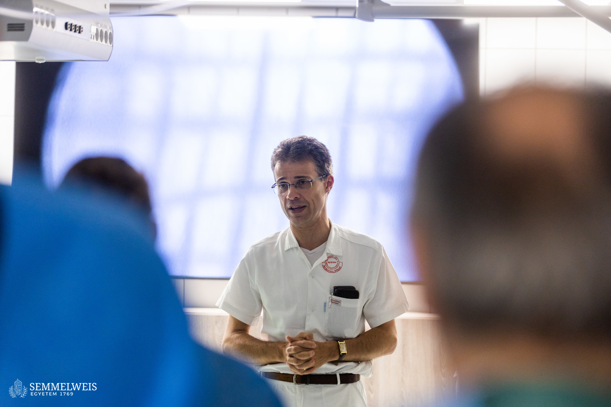
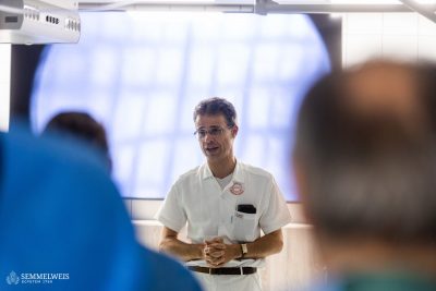 “It was a long-time dream come true and an important milestone in education, training and research when the Interdisciplinary Cadaveric Operating Room and the refurbished mortuary department were completed last November. Now, at last, we no longer need to take the expertise and experience accumulated at the Department of Anatomy, Histology and Embryology abroad for training, but rather can organize courses locally to enrich the anatomical knowledge of clinicians such as specialists, residents and trainee specialists. Courses launched almost immediately after the handover of the facilities, but the gynecological cadaveric surgery training course organized in early June is regarded the most outstanding of all,” said Dr. Alán Alpár of the Department of Anatomy, Histology and Embryology. “The June course was remarkable not only for the number of participants. The very favorable participant-instructor ratio enabled continuous consultation between participants and instructors, with twelve participants practicing at four tables, each table with an expert and an anatomy specialist facilitating the training. As an additional novelty, the new fixation method developed by the department staff allowed the participants to work with the endoscope on cadavers in life-like environment with soft, warm tissues,” underlined Dr. Alán Alpár.
The occupancy rate of the Interdisciplinary Operating Room is increasing, as several other specific training courses preceded the laparoscopic gynecological surgery training, and seven more courses are scheduled for later this year. “Professional and further training is an indispensable part of the profile of an anatomy department, and this was one of the priorities of my tender for the post of director, along with opening up anatomy education to clinical and postgraduate medical training. Our objective is to keep supporting students and professionals even beyond basic anatomy education, to involve clinicians in theoretical education and to have theoreticians lecture to clinicians”, concluded Dr. Alán Alpár, noting that reaching this goal will increase the university’s embeddedness and will also improve its position in international rankings.
In the Interdisciplinary Cadaveric Operating Room of the Department of Anatomy, Histology and Embryology, procedures are performed on deceased persons who offered their bodies for medical school education and research during their lifetime or in the absence of their denial, through a declaration of consent by an authorized next of kin following death. During the training, participants can improve the necessary manual skills on these cadavers without compromising patient safety.
Anita Szepesi
Translation: Judit Szabados-Dőtsch
Photo: Bálint Barta — Semmelweis University
