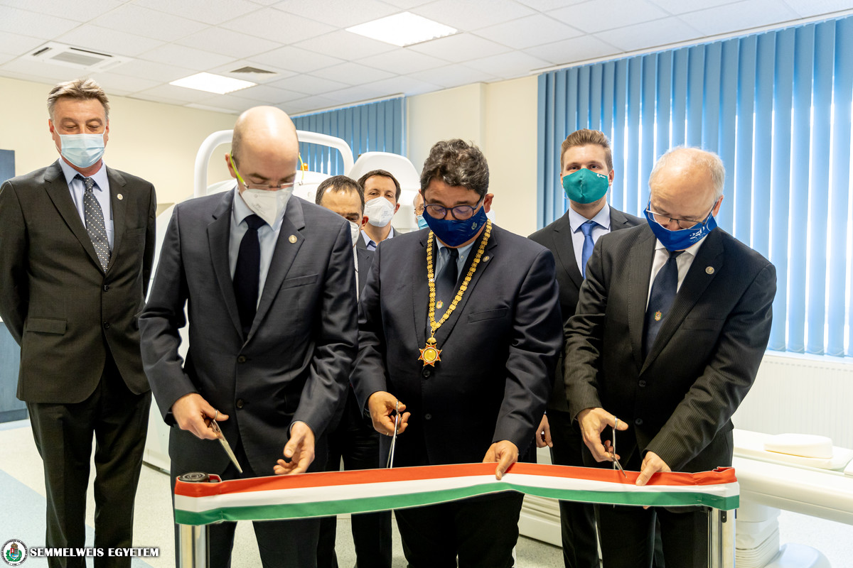
A new AnyScan™ PET-CT device, acquired in the framework of the VEKOP tender, was handed over to Semmelweis University at a ceremonial ribbon cutting. Thanks to the development of more than 400 million HUF, it is now possible to map incurable diseases and their pathological factors, such as aggressive and malignant tumors, cardiovascular diseases and circulatory disorders of the central nervous system. The high-sensitivity and high-resolution imaging equipment enables functional imaging and, as a result of the collaboration with the manufacturer, it also includes a three-detector SPECT module that is unique in the world.
The university won over 400 million HUF from the European Union and Hungarian funds in the framework of the Competitive Central Hungary Operational Program (VEKOP). This project enabled the procurement of an AnyScan™ PET-CT device and the establishment of the special environment. The “Innovative large animal models for clinical therapeutic research” project was implemented at the Department of Nuclear Medicine of the Medical Imaging Center under the leadership of the Department of Biophysics and Radiation Biology.
“The development and the project are outstanding examples of translational research with results that may be used directly by the patient bed”, pointed out Dr. Béla Merkely, Rector during the ceremony that was held in compliance with the current epidemiological measures.
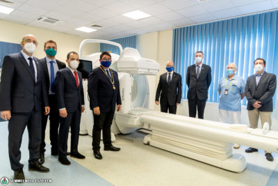
Thanks to the integration of three different imaging technologies, SPECT/PET/CT, in one device, the most complex scientific questions may be answered. During examinations, the function and the smallest details are visible, which enable the translation of new therapeutic products and diagnostic procedures from pre-clinical trials into human use.
 “A dream of more than 10 years has come true, as this device completes Semmelweis University’s “From Molecule to Man” translational research line”, pointed out Dr. Miklós Kellermayer, project leader, Director of the Department of Biophysics and Radiation Biology, Dean of the Faculty of Medicine.
According to the dean, now they have the opportunity to study the problem encountered by the patient bed on the level of individual molecules, while following the processes in small and large animal models, as well as in humans, and finally use the results by the patient bed again. Dr. Miklós Kellermayer also said that this approach is implemented in their current study related to the coronavirus, when the examination of the problem was narrowed down to the level of individual viral particles in order to understand the nature of the pathogen and the pathomechanism of the disease.
“The project serves our long-term goal of obtaining the most diverse, spatially and temporally related information about the smallest possible element of the living human body, thus enabling us to understand more precisely the normal and pathological processes in the living human body”, Dr. Miklós Kellermayer said.
In addition to the Department of Biophysics and Radiation Biology and the Department of Nuclear Medicine, several other university institutes, including the Department of Urology, the Department of Neurology and the Department of Surgical Research and Techniques are also involved in the specific research already planned for the project.
The new infrastructure with high sensitivity on molecular level enables the study of large animal models, thus contributes to the extensive mapping of incurable diseases and their pathogens. The main areas of research include cardiovascular and cerebrovascular diseases, aggressive and malignant tumors, and the latest pharmacons can be tested.
 “The new infrastructure will help to create real interdisciplinary cooperation between the institutes. The completion of the project coincided with the establishment of the unified Medical Imaging Center, therefore we can extend the spectrum of clinical research, and strengthen cooperation between different disciplines (for example nuclear medicine, radiology)”, highlighted Dr. Pál Maurovich-Horvat, Director of the Medical Imaging Center.
He also emphasized that the project is a good example of the collaboration between industry and the university that allows the immediate and continuous update of the equipment by the latest developments.
Dr. Tamás Györke, Director of the Department of Nuclear Medicine, reminded that the current infrastructure is one of the most outstanding developments of nuclear medicine at the university in the last decade. The first SPECT-CT was installed in 2011, which was the first combined modality at the university. The next major step was the introduction of PET-CT for human examinations at the end of 2016.
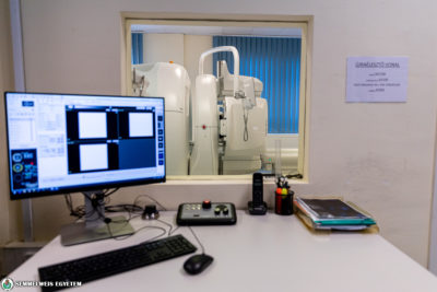
He emphasized the importance of the fact that the current new device may primarily serve research, for which the existence of the three modalities is particularly advantageous, but at the same time it is also suitable for diagnostic testing in the context of patient care.
 The project made it possible to procure PET and CT modules, but also added a SPECT component as part of the research and development cooperation with the equipment manufacturer, Mediso Kft.
“It is unique even on a global level that all three modalities are combined in this high-end AnyScan™ TRIO SPECT/CT/PET device, and the three SPECT detectors are complemented by a so-called Multi-PinHole collimator. This technology allows SPECT to use imaging technology and to produce image quality similar to those of PET. Furthermore, the project is another example of the close partnership between Mediso and Semmelweis University”, pointed out Gergő Bagaméri, product development director of the company’s preclinical business.
“In addition to medical research, the SPECT modality also provides an opportunity for significant research and developement activities in engineering, physics and industry related areas, therefore a dynamic SPECT test method will be developed with the cooperation of Semmelweis University, the industrial partner (Mediso Kft.), Budapest University of Technology and Economics and Eötvös Loránd University. The development aims to achieve better image quality by developing new imaging techniques.”, emphasized Dr. Béla Kári, research fellow, engineering physicist of the Department of Nuclear Medicine.
In order to support a wide range of research areas, it is also planned to allow the public use of the research laboratory for the study of large animal models as a “public treasure”. This means that it would operate as a core laboratory and the research infrastructure will be available for the entire university on the basis of appropriate regulations, and it will also be available to Hungarian companies and research sites.
Pálma Dobozi
Photo: Attila Kovács – Semmelweis University
Translation: Katalin Illés-Romhányi