What causes pain associated with cartilage wear?
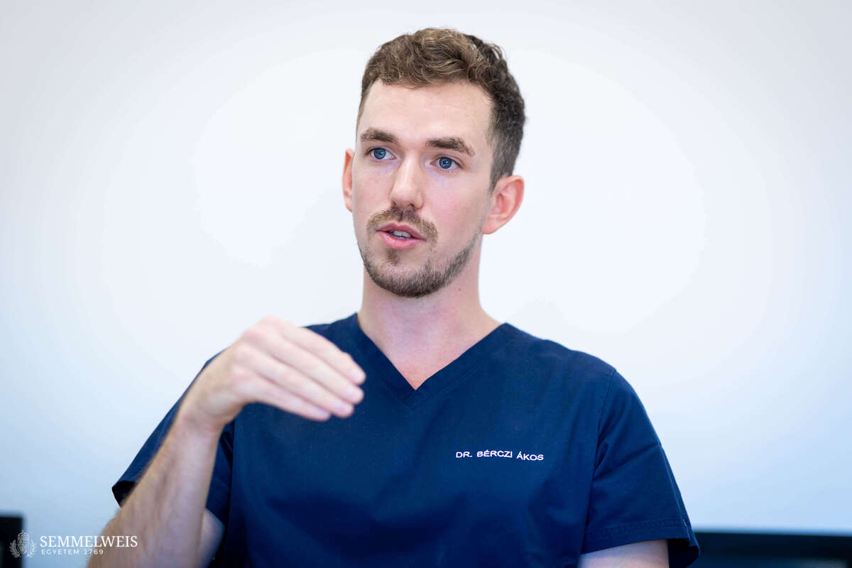 “At least one million people in Hungary suffer from knee osteoarthritis – commonly referred to as cartilage wear – and the chronic pain resulting from it. When we speak of knee osteoarthritis, we generally think of the wear and thinning of the hyaline cartilage covering the surfaces of the bones that form the joint; however, the pain caused by knee osteoarthritis results from the combined presence of a complex set of five to six factors,” explained Dr. Ákos Bérczi, consultant at the Department of Interventional Radiology, in an interview with our website. “In addition to cartilage thinning, inflammation of the synovium – the membrane lining the joint – and the broader range of inflammatory mechanisms play a particularly important role in the development of pain, along with cellular processes associated with aging, as we pointed out in our recently published paper co-authored with colleagues from Semmelweis University and international researchers,” emphasized Dr. Ákos Bérczi.
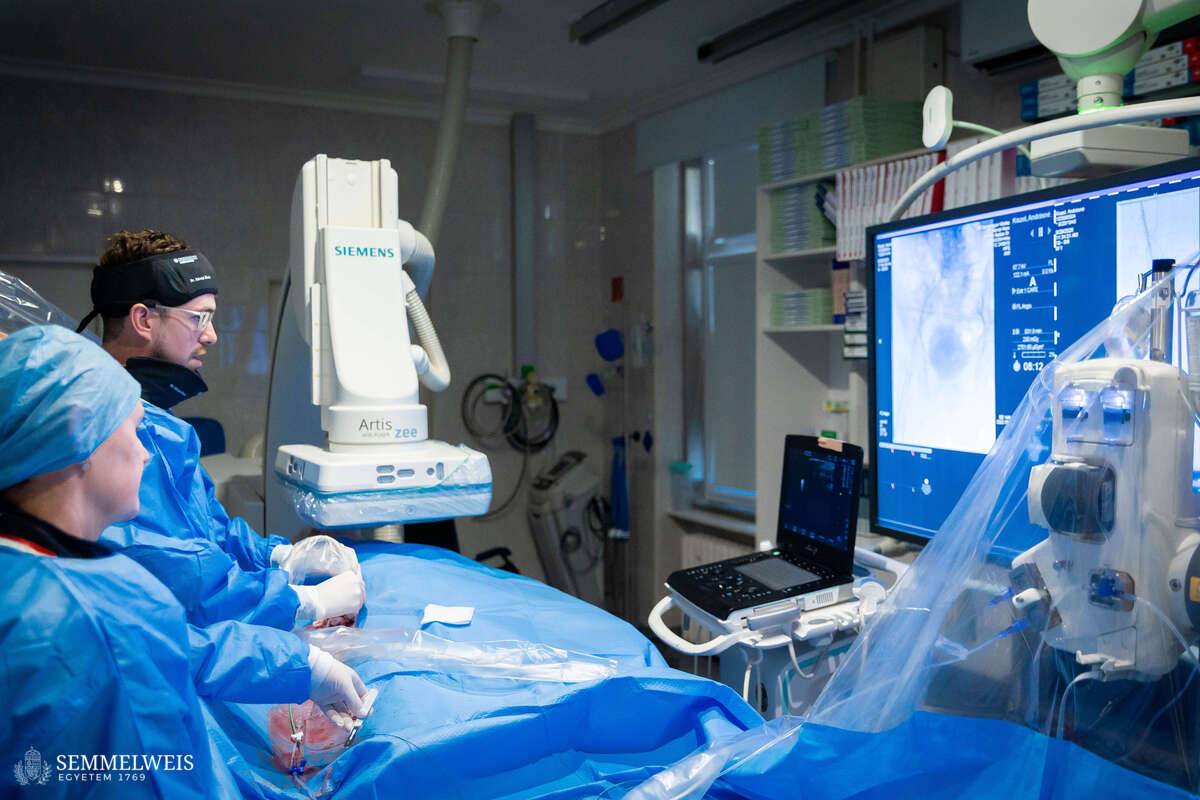
The GAE procedure used at Semmelweis University does not target the cartilage itself, but rather the inflammatory neovascularization associated with knee osteoarthritis. Besides the tiny blood vessels that proliferate abnormally in the inflamed synovium, pain-sensing nerve fibers may also appear, meaning that the vascular and inflammatory processes can contribute to persistent chronic pain. During the procedure, the interventional radiologist uses a microcatheter to target these areas with abnormally increased blood flow. The success of the therapy largely depends on selecting suitable patients.
Not an alternative to knee replacement
This method is primarily intended to provide relief to patients who have been suffering from pain associated with knee osteoarthritis for at least three months and who continue to experience significant symptoms despite appropriate conservative treatment – such as lifestyle changes, weight loss, physical therapy, pain or anti-inflammatory treatment, or, in some cases, intra-articular injection therapy. “During patient selection, it is important to rule out conditions that require different treatment and to assess the condition of the lower extremity vascular system. An MRI scan can help evaluate the nature of osteoarthritis, inflammation of the synovium, and other abnormalities that may explain the pain,” added Dr. Alexisz Panajotu, consultant from Diagnostic Imaging at the Városmajor Heart and Vascular Center. The therapy can be performed successfully in patients typically between the ages of 40 and 80 who do not yet require knee replacement surgery as GAE is not an alternative to knee replacement; for some patients, it may offer a new option in the therapeutic gap between conservative treatment and surgical intervention.
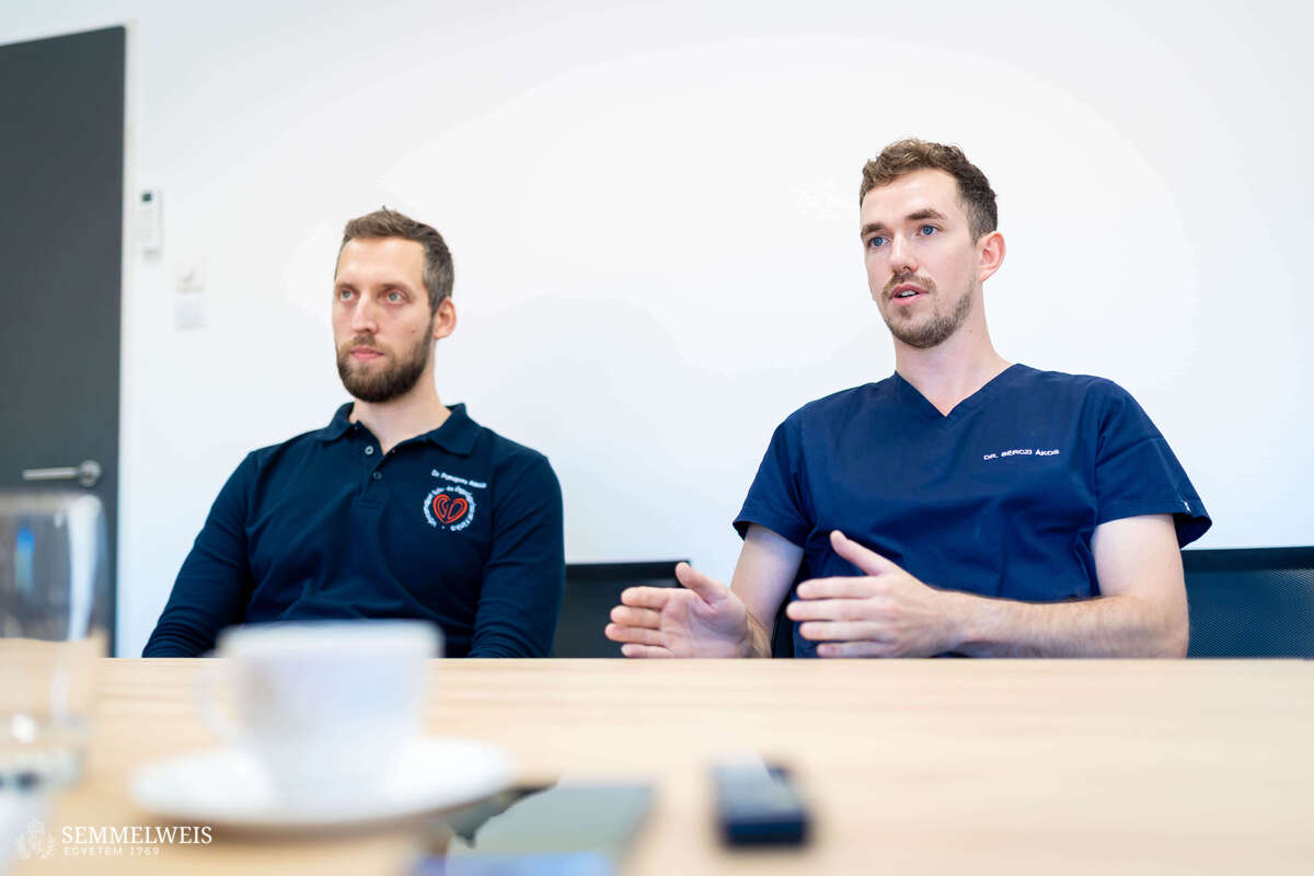
The method, first described by Yuji Okuno and his colleagues for the treatment of knee osteoarthritis in Japan in 2014–2015, has been adopted by an increasing number of centers in Europe and North America in recent years. At the Városmajor Heart and Vascular Center, work to identify this patient group began a year ago. “Initially, we performed the first procedures in collaboration with an expert from England – Dr. Mark Little, an interventional radiologist and internationally recognized expert in GAE – and a multidisciplinary university team consisting of rheumatologists, orthopedic surgeons, as well as diagnostic and interventional radiologists,” recalled Dr. Ákos Bérczi. Since the first procedure in February, 22 patients have been treated using this method, and in line with the international literature, early follow-up revealed a measurable reduction in pain in nearly 80 percent of patients compared to their pre-procedure condition.
Patients can be referred from anywhere in Hungary
Semmelweis University currently operates one of the largest structured GAE programs in Hungary. Patients can be referred to the Városmajor Heart and Vascular Center by a rheumatologist or orthopedic surgeon from anywhere in the country, and those who, upon completion of their examinations, are deemed eligible for a procedure financed by NEAK, will typically undergo an intervention requiring one night of hospital observation. Clinical improvement may then develop gradually.
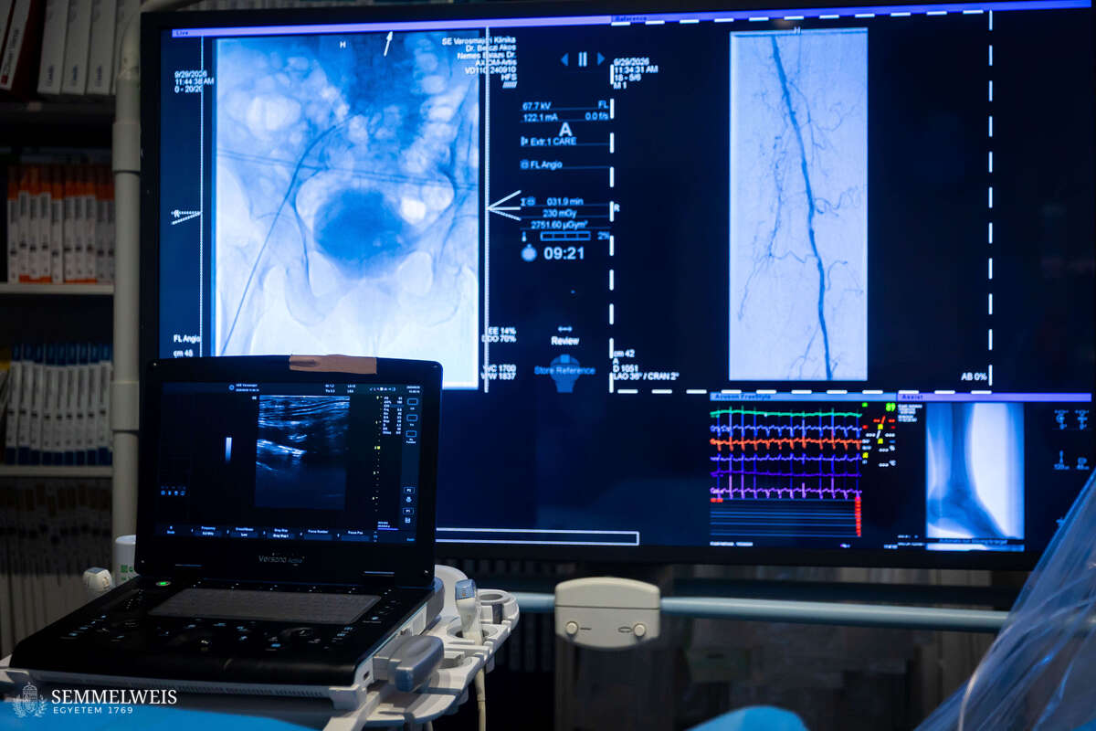
“The patient pathway from the Department of Rheumatology and Immunology is already in place, and one is currently being established for the Department of Orthopedics as well,” stated Dr. Ákos Bérczi. “We have sufficient capacity to care for incoming patients, and we are able to admit patients on a weekly basis,” he added. As he noted, research is still ongoing regarding whether this procedure – which is primarily aimed at reducing pain through anti-inflammatory treatment – also slows down the degenerative process itself.
About the program
The genicular artery embolization program is being developed through multidisciplinary collaboration, with the support of Dr. Béla Merkely, Rector of Semmelweis University and Director of the Városmajor Heart and Vascular Center. The interventional radiology-related professional and scientific work is coordinated by Dr. Edit Dósa, Head of the Department of Interventional Radiology, and Dr. Ákos Bérczi. Also participating in the clinical and scientific program are Dr. Alexisz Panajotu, radiology resident Dr. Fanni Szablics, and medical student Dóra Hámori. Dr. György Nagy, Director of the Department of Rheumatology and Immunology, provides the rheumatological expertise for the program. Representing the Department of Orthopedics in this collaboration are Deputy Director Dr. Gábor Skaliczki as well as residents Dr. Bence Stubnya and Dr. Péter László Rein.
Anita Szepesi
Translation: Dr. Balázs Csizmadia
Photos by Boglárka Zellei – Semmelweis University