The development significantly expands diagnostic capacity in these areas. Pediatric patients currently account for every second MRI examination performed at the university. Each year, hundreds of children with hypoxic brain injury, central nervous system disorders, or cancer require MRI scans. The new system is capable of examining up to 300 patients per month, from neonates to adolescents.
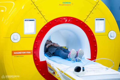
The scanner offers high-resolution tissue imaging combined with AI-based motion correction, enabling precise examinations even when a child moves during the procedure. Its wider bore and reduced noise levels are designed to ease examination-related anxiety and may help reduce the need for anesthesia.
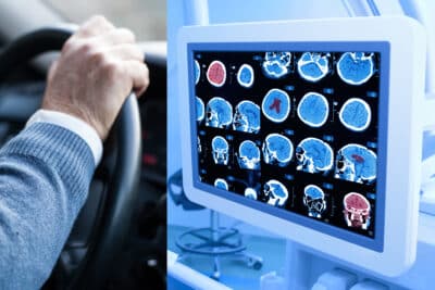
In pediatric oncology, MRI plays a central role in both diagnosis and therapeutic follow-up, allowing detailed assessment of tumor structure, extent, relationship to adjacent organs, and potential recurrence.
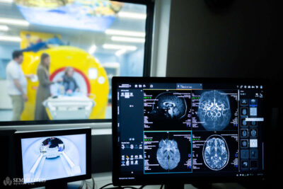
The new Siemens MAGNETOM Vida 3T system is also expected to make a meaningful contribution to research. Its high image quality and reproducibility make it well suited for prospective clinical and imaging studies, particularly in pediatric oncology, neurology, and neurodevelopmental research, where standardized imaging is essential for reliable longitudinal assessment. The system will additionally support the development and validation of new pediatric MRI protocols.
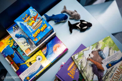
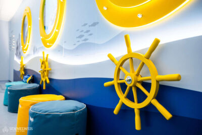
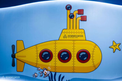
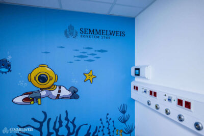
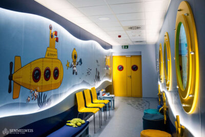
Complementing the technological upgrade, the waiting and examination areas have been redesigned with a submarine-themed, child-centered interior, aimed at reducing anxiety and encouraging cooperation during imaging procedures.
Gallery


Photos by Bálint Barta – Semmelweis University