Diabetes, tumors, central and peripheral nerve problems, facial nerve palsies, viral infections, multiple sclerosis, and even congenital brain diseases can lead to corneal nerve lesions that cause the ocular surface to lose its ability to perceive. This means, for example, that the eye does not automatically water or close when an insect flies into it.
 To remedy this, Dr. Zoltán Klárik, a plastic surgeon at the Department of Surgery, Transplantation and Gastroenterology, has introduced a special procedure at Semmelweis University, the presentation of which he saw at the annual meeting of the American Society for Reconstructive Microsurgery. “This technique is used for pathographies where the insensitivity of the ocular surface prevents the patient from feeling any damage to the cornea, that is, the eye’s defense mechanism is inadequate. As a result, the patient may lose the blink reflex, tears do not wash over the eye so that it is subject to constant irritation. In addition, there is a lack of the proteins that are produced by the nerves, and which are responsible for the integrity and regeneration of the corneal epithelium. This can lead to ulcers forming on the eye, which can eventually cause blindness,” Dr. Zoltán Klárik explained.
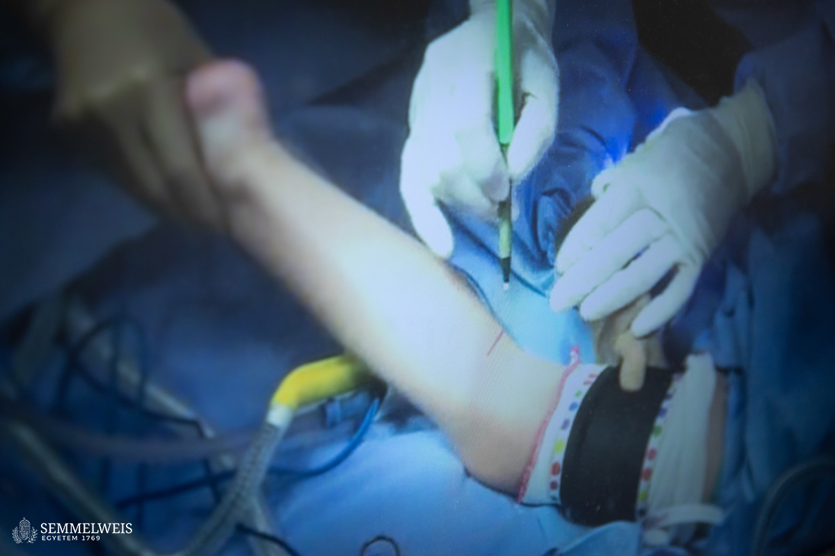
The first operation was performed on a middle-aged woman by a team of surgeons and ophthalmologists. During the special procedure, a 15-centimeter nerve fiber was removed from the patient’s lower leg in a way that did not cause any functional deterioration in the leg. As the plastic surgeon pointed out:
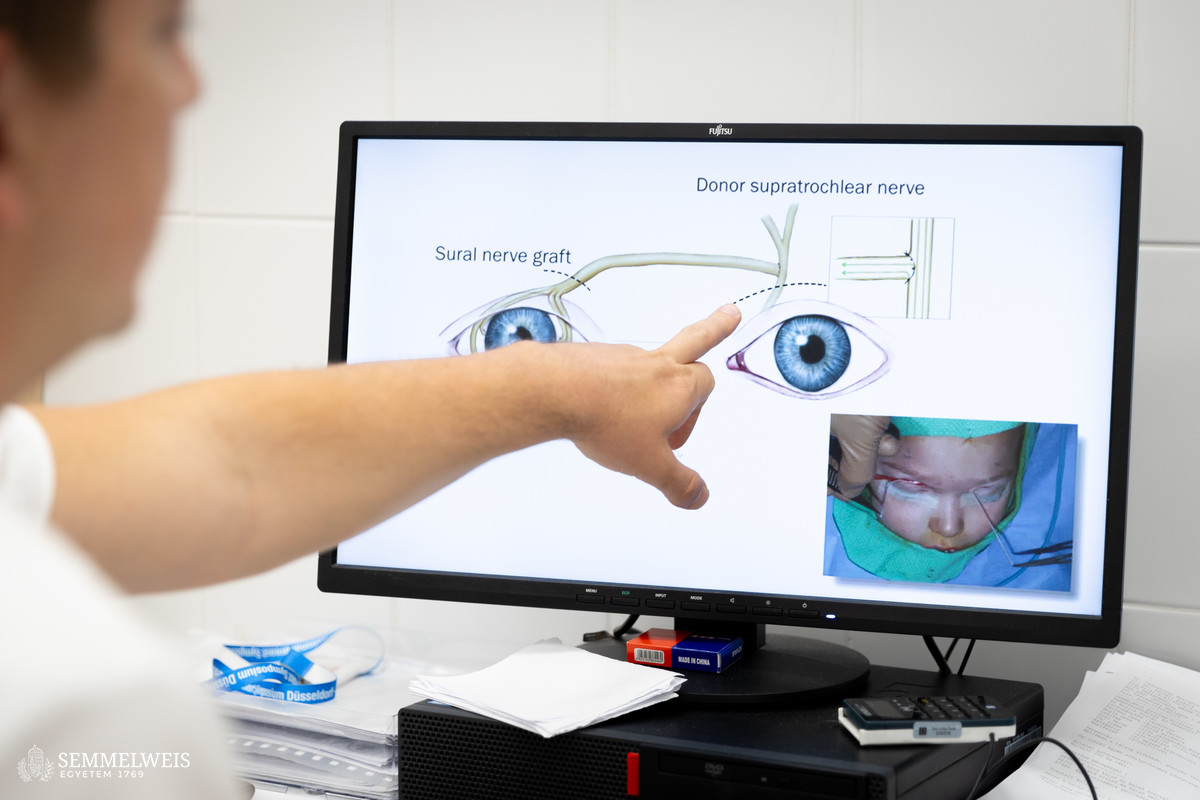
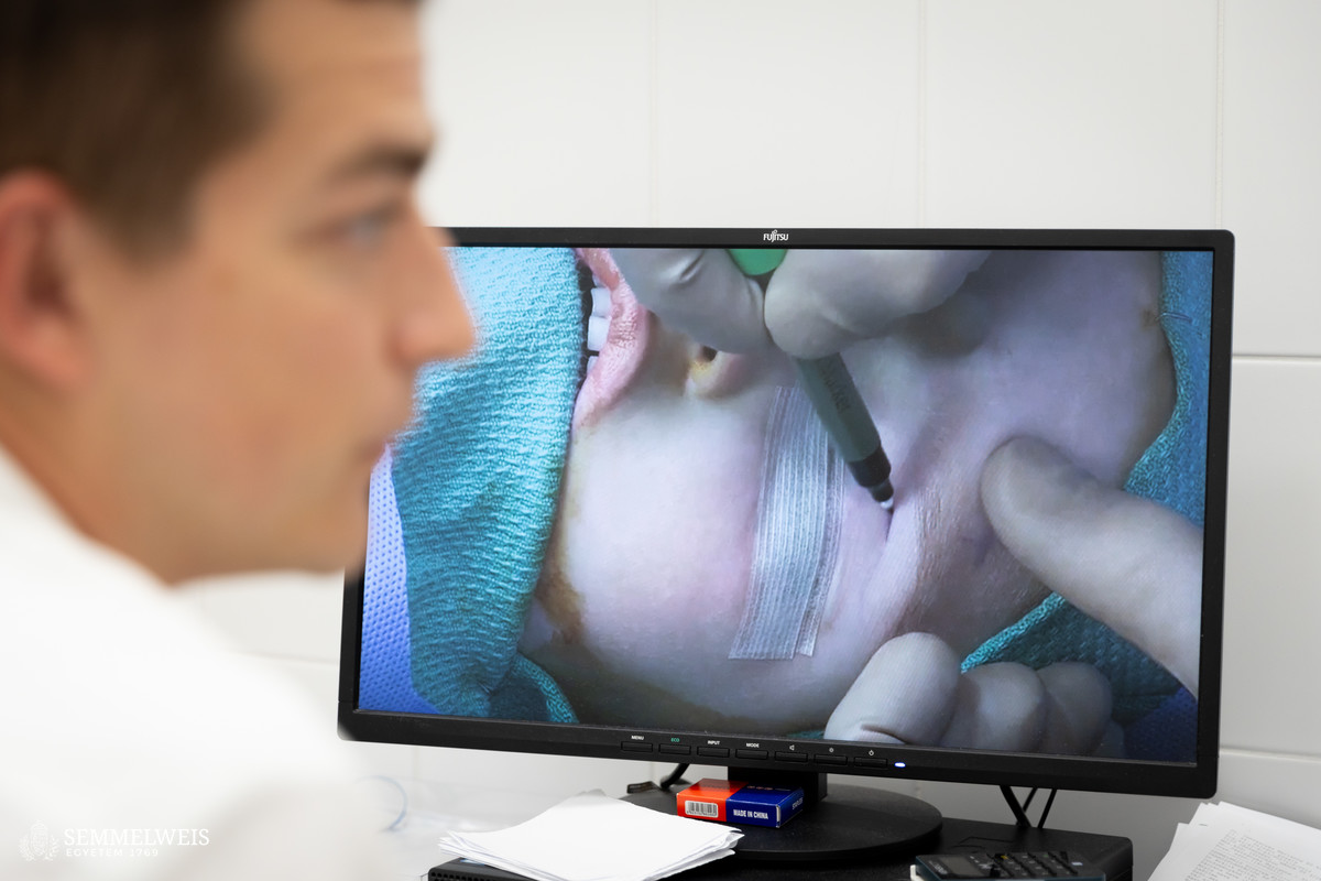
My task was to divide the thick nerve taken from the lower leg into several fibers, suture them under a microscope, and then guide the nerve from the intact side through a so-called subcutaneous tunnel system formed at the forehead.
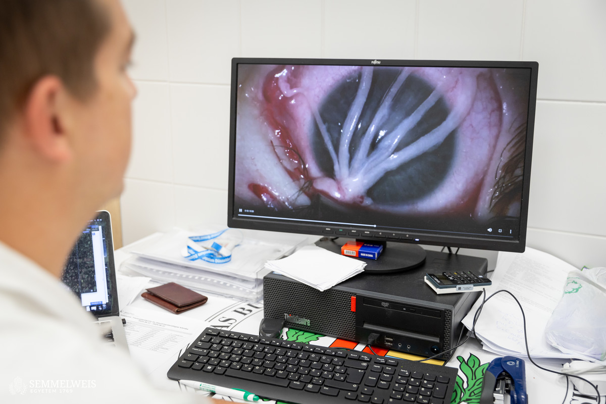
 The ophthalmology team, led by Dr. Ágnes Füst, then inserted the nerve through the eyelid into the ocular surface and guided it under the conjunctiva to fix it in place at five points around the cornea. During the specialized ophthalmic and plastic microsurgery procedure, doctors sutured the millimeter-thick structures under an operating microscope. “The most difficult part of the operation was organizing the sequence of the steps so that the plastic surgeon and the ophthalmology experts could work alternately,” said Assistant Professor Dr. Ágnes Füst, recalling the difficulties of the four-hour procedure performed at the Department of Ophthalmology.
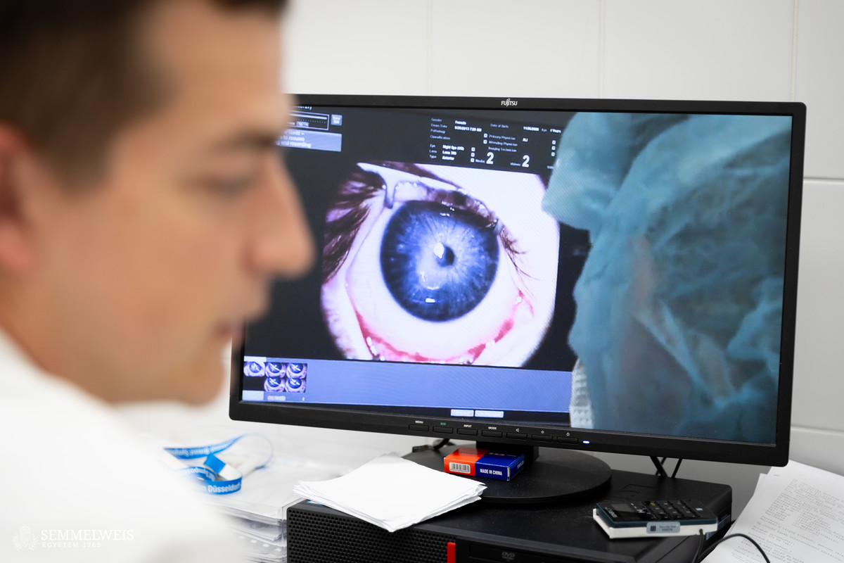
The patient had been suffering from recurrent, non-healing corneal ulcers due to nerve damage caused by a central nervous system tumor. She had undergone three eye surgeries previously to achieve healing of the ulcers, the epithelization of the cornea, and to prevent further deterioration of vision. Since the surgery, however, the ulcer has not returned and the patient’s quality of life is constantly improving; thus, the team was able to prevent her from losing her vision completely.
Gallery
Orsolya Dávid
Translation: Dr. Balázs Csizmadia
Photos by Boglárka Zellei – Semmelweis University