Since 2017, a team of about 10 researchers from the Institute of Anatomy has been investigating the neuronal explanation of schizophrenia in collaboration with the American, Danish and Croatian university. Although there have been numerous studies on the disorder, which was described 110 years ago, the exact neuropathology of the disease remains unexplored.
Our aim was to identify the exact neuron populations and receptors that could at least partly explain the symptoms of the disease,
Dr. István Adorján, a research fellow at the Department of Anatomy, Histology and Embryology explained to our website.
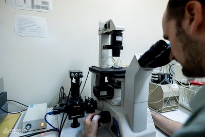
A special feature of their studies was that they analyzed post mortem samples from the same donors at different biological levels. We performed single-cell RNA sequencing of a total of 220,000 neurons at the single-cell level, with morphological and topographical analysis of 120,000 neurons, and detected the changes in cell shape and location in the dorsolateral prefrontal cortex,” Dr. István Adorján said. “We investigated at the cellular level, the University of Copenhagen did the transcriptomic studies, the Harvard researchers excelled in data analysis, and the Zagreb researchers performed verification experiments to target the identified mRNAs and cell populations with multiplexed signaling,” he added.
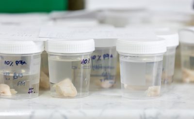
“In the experiment, samples from 10 schizophrenic and 10 control brains were analyzed, with samples requested from different European brain banks, most of which were obtained by the Hungarian team. This posed a logistical, methodological and conceptual challenge,” the team leader pointed out.
“The quality of the samples is crucial, and single-cell sequencing is very sensitive to the time between death and freezing. It was also essential to have both frozen and formalin-fixed samples from the same brain, which is rare because not all brain banks use both methods,” he emphasized. For immunohistochemical detection at the cellular level, formalin-fixed samples are needed for immunohistochemistry, while frozen samples are needed for mRNA sequencing.
“My basic hypothesis was that certain inhibitory neuron populations, which have been less studied, are involved in schizophrenia. The question was whether this was supported by foreign collaborators working with single-cell sequencing,” Dr. István Adorján explained. The study confirmed the hypothesis, revealing the same cell populations by single-cell sequencing.
“According to the study, the inhibitory neurons located in the upper layer of the cortex-calretitin positive neurons and parbaalbumin neurons – are the most affected, but we were able to classify more than a dozen inhibitory neurons. This technique is also useful to discover new neuron populations and to describe receptor sets that can be used to subsequently manipulate these neuron populations and to target them as therapeutic targets for drug development,” the research leader added.
It is estimated that between 70 and 80 million people worldwide have schizophrenia, with the number of people with autism estimated at between 40 and 50 million. The high and consistent prevalence of the disorder – 1.1 per cent prevalence in schizophrenia and 0.6 per cent in autism – suggests that evolutionary drivers are likely to be behind both syndromes, the researcher points out.
“It is not yet known which cells are affected by current drug therapies or which neuronal populations populate the subcortical areas. Until this is accurately mapped, the chances of detecting altered cell populations in pathologies are minimal. We are working to map the composition of the different brain areas, compare the control brain with samples from schizophrenic and autistic donors (our research also covers the neuropathological background of autism) and draw conclusions from the difference between the two as to which cell populations may be altered,” Dr. István Adorján explained.
Our study shows that there is a population of cells that has received less research attention, namely the special inhibitory and excitatory neurons, which are mainly located in the upper layers of the cortex and which are damaged. The importance of this so-called dorsolateral prefrontal layer in both diseases has been described previously, and MRI scans have shown that these cortical areas function differently in affected patients, but what exactly underlies this has not been known. Indeed, the detection of altered cell populations is already the domain of neurohistology and neuropathology, and our study was the first in which we were able to correlate the results obtained by single-cell sequencing with those obtained by neurohistological methods, and they pointed in the same direction. Furthermore, the significance of our research lies in the combination of neurohistological and transcriptomic methods. This was the first time we have worked with such a complex approach and sample, filling a gap of several decades,” the researcher from the Institute of Anatomy, Tissue and Developmental Sciences said. He added that work will continue until the composition of these areas is fully understood.
“Our aim is to use the results in applied research, to study these neuron populations in animal models, in in vitro stem cell experiments and to influence the truly altered ones in different ways, but this is already the field of drug development,” Dr. István Adorján emphasized.
In the lucky case, our research could lead to a new treatment for schizophrenia,
the researcher added.
Dr. István Adorján is a research physician at the Department of Anatomy, Histology and Embryology at Semmelweis University. He graduated from the Faculty of Medicine, Semmelweis University in 2005 and from the János Szentágothai Doctoral School of Theoretical Neurosciences in 2012. He was a postdoctoral fellow at the Oxford Brain Bank, University of Oxford from 2013-2016. In 2021 he was awarded the János Bolyai Research Scholarship of the Hungarian Academy of Sciences.
Anita Szepesi
Translated by Rita Kónya
Photo: Attila Kovács – Semmelweis University; wildpixel (iStock) – illustration


