Using lower dose X-ray and less contrast agent, a new device provides better image of the vascular system than before. The device was developed by a start-up company of Semmelweis University and it has already been used on a daily basis at the Városmajor Heart and Vascular Centre in interventions related to peripheral vascular diseases, such as in the diagnosis of vasoconstriction or vasodilator therapies. The Centre is the company’s primary collaborative partner. Kinepict has recently won € 1.5 million at the most prestigious innovation competition of the European Union.
 Cardiovascular diseases are a leading cause of death worldwide, circulatory disorders account for half of all deaths in Hungary. Approximately 500,000 people are affected by abnormalities of the vascular system (eg. vasoconstriction, atherosclerosis), which can be detected by angiography. The invention of Kinepict, a start-up company of Semmelweis University revolutionizes this traditional imaging technology using contrast agent.
“Thanks to the technology developed by the start-up company, image quality has been enhanced, the exposure of patients and staff to X-ray radiation has been reduced by 70% according to clinical studies, while the amount of contrast agent used in the procedure has been decreased by 50%, which also reduces adverse effects on the patient.”, said Dr. Krisztián Szigeti and Dr. Szabolcs Osváth, founders of Kinepict and senior research fellows at Semmelweis University’s Department of Biophysics and Radiation Biology.
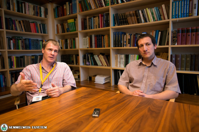 “X-ray images of blood vessels have been processed in the same way for 30-40 years and the new software has transformed this practice by changing the mathematical evaluation of the images.”, said Dr. Szabolcs Osváth.
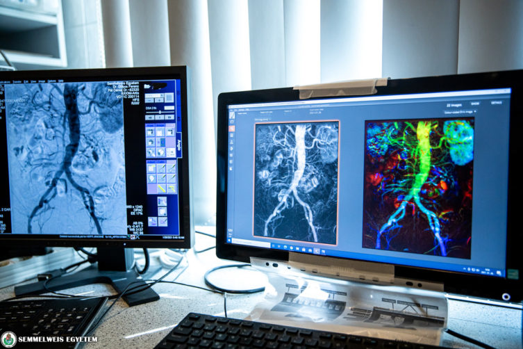
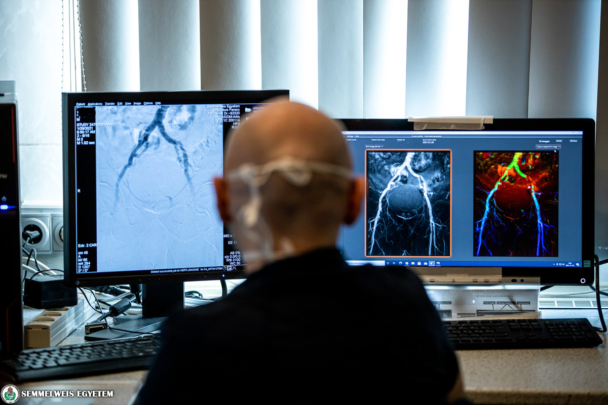
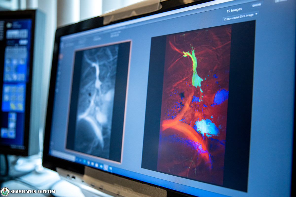
The technology is based on kinetic imaging, which involves data collection and data analysis of several consecutive X-ray images. The new image processing technology is called digital variance angiography (DVA), which enables the extraction of more information from tests using X-ray.
“As part of the EIC Accelerator grant application, the start-up has recently won an EU grant of € 1.5 million for the further development of the innovation, the software and the protocol, as well as for its dissemination. Kinepict is the first Hungarian company to win the grant in the field of healthcare. We were selected as one of the 32 winning projects out of 4,200 European companies.” said Dr. Krisztián Szigeti.
The project provides an opportunity to extend scientific and research collaborations as well. In this field, the company’s primary partner is Semmelweis University, which originates from a cooperation with the Városmajor Heart and Vascular Centre. This joint project revealed that the new technology can primarily be used in the field of vascular medicine.
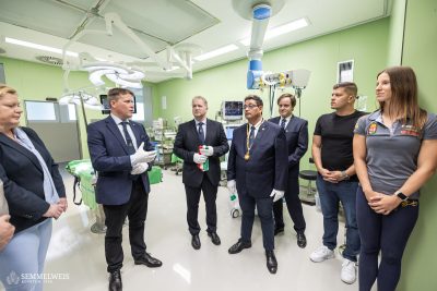
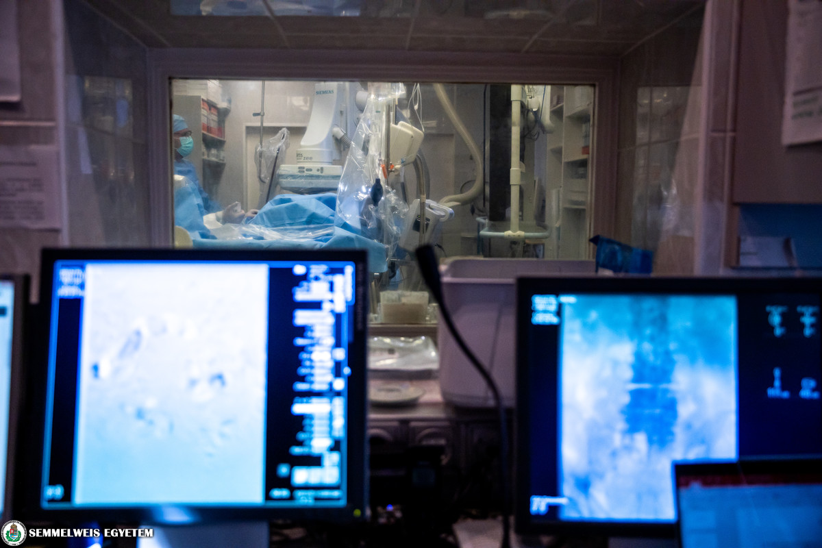
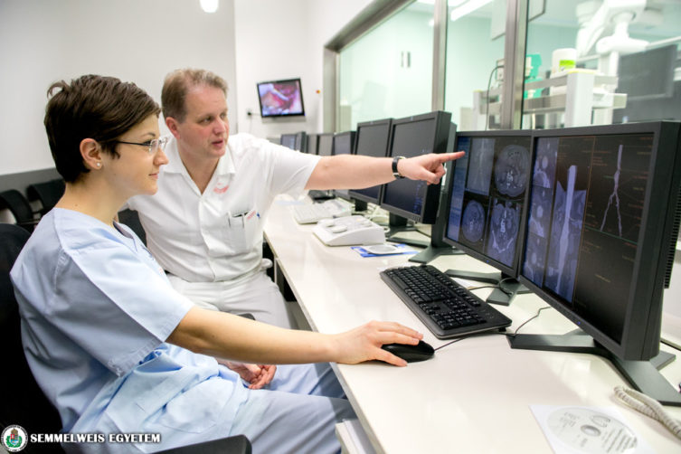 “The new technology is already in use in angiography on a daily basis at the Városmajor Heart and Vascular Centre. The software is used in the most common peripheral interventions, in diagnostic procedures and in catheter surgeries (eg. the examination of vasoconstrictions in the neck and lower limbs, vasodilation procedures). It is very important for angiologists to obtain good quality images with as little radiation exposure and contrast agent as possible. This technology makes it possible.” said Dr. Péter Sótonyi, head of the Department of Vascular Surgery and Endovascular Medicine.
The new imaging processing technology enables the safe examination of blood vessels in patients with renal insufficiency because instead of the traditional iodine contrast agent, which requires extreme precautions in case of this patient group, the use of carbon dioxide also produces good quality images of the blood vessels. This innovation also helps a lot of people to take their first steps in their scientific career, as the first PhD dissertation defence has already been presented on the analysis of the efficiency of the technology in the imaging examination of lower limb blood vessels.
Gallery
Kinepict software, as a medical device is licensed in Europe and in the USA. In addition to the Heart and Vascular Centre, it is used in Kecskemét in Hungary. There are several scientific collaborations with Austrian and German labs where the device is used in research and in health care as well. The software is compatible with any kind of angiographic imaging system and it can be added to older devices. The goal of Dr. Krisztián Szigeti and Dr. Szabolcs Osváth is to make the software better known in the field of angiology and to prove its efficiency in as many indications and patient groups as possible.
Patented algorithm
In 2D X-ray angiography the use of DSA [digital subtraction angiography] is the standard protocol, which captures a series of images containing contrast agent and then extracts an image (the so-called “mask”) from the other images. These extracted images make up a DSA video and their proper integration renders a DSA image. In contrast, DVA, the new technology uses all the images of the series without extraction and calculates the scattering intensity for every single pixel. The patented algorithm highlights functional and movement related information (i.e. the flow of the contrast agent), while suppressing the noise, which significantly enhances image quality compared to that of the DSA. This was also confirmed by three completed clinical trials and according to statistical evaluations and the observations of specialists, DVA produced better quality images than the traditional DSA method.
Pálma Dobozi
Photo: Attila Kovács – Semmelweis University
Translation: Ágnes Raubinek