The research group led by Dr. Botond Roska have succeeded in creating an artificial retina that functions like the living human organ in a healthy adult. The research results published in Cell suggest that the so-called retina organoid may open up possibilities to develop personalised therapies targeting vision. Authors from Semmelweis University also contributed to the publication.
The retina is a nerve tissue in the eye responsible for sensing light and organizing visual information. Although it seems to be an easily accessible organ thanks to its location, its cellular functions have not yet been mapped in detail. Most knowledge about vision is based on animal experiments. As Dr. Arnold Szabó stated in connection with a previous Science publication, it has been proven that conclusions drawn from animal experiment results cannot be directly applied to humans. The study of the human retina is rendered difficult by the tissue’s sensitivity; the tissue removed from the body suffers irreversible damages in a few minutes as oxygen and nutrients supply are suspended. The team of the Institute for Molecular and Clinical Ophthalmology Basel headed by Dr. Botond Roska have managed to artificially generate light-sensitive retina organoids from peripheral tissues (connective tissues and blood) that basically correspond to the properties of the human retina. The procedure used cells that have already undergone differentiation. These cells were isolated from the skin or blood of adult donors, which were turned back into stem cells. These stem cells were then used to grow an unlimited number of retina tissues outside the body, which corresponded to the original organ.
In addition, the researchers have been the first to measure light response with electrophysiological methods in isolated human retina of organ donors that may bring us closer to understanding retina’s function. The retina samples from organ donors and the cultured retina organoids also underwent unicellular transcriptome analysis. Using the molecular biological method, the gene activity of thousands of cells were studied on cellular level and based on the results the cells were organized into groups, called clusters. The retina tissue obtained from donors and the cultured tissue showed high correlation regarding the pattern of the cells’ genetic activity. Moreover, cellular types playing key roles in several genetically determined ophthalmic diseases have been identified.
 The discovery was preceded by several years of research, involving the Retina Laboratory of Semmelweis University’s Department of Anatomy, Hisotlogy and Embryology. Dr. Arnold Szabó, head of the laboratory and assistant professor at Semmelweis University and Dr. Ákos Kusnyerik, research fellow at the Department of Ophthalmology are among the authors of the publication as contributors. According to Dr. Arnold Szabó the research results suggest that in the future personalised therapy may be available for patients, as the properties of the organoid cultured from the patient’s cell carry the patient’s disease as well. Thus, the effects of potential therapies may be directly studied in the artificial retina cultured in a dish.
The publication is available on the following link:
https://www.sciencedirect.com/science/article/pii/S0092867420310047
Ádám Szabó
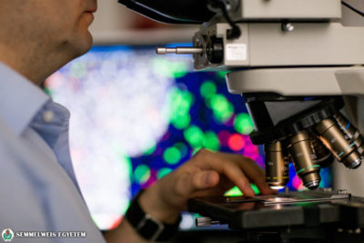
Photo: Attila Kovács – Semmelweis University
Illustration (retina organoid): iob.ch
Translation: Ágnes Raubinek