Semmelweis University’s Medical Imaging Center (OKK) participated in a unique research project in Hungary, where the best-preserved of the ten Roman-era mummies found in Hungary to date was examined. “As part of the collaboration between the MTA–ELTE HTK Momentum Bioarchaeology Research Group and the university, we have previously examined Roman-era bone finds using photon-counting detector CT, but this is the first time we have been able to do so with a mummy find,” Dr. Ádám Békésy-Szabó, radiologist and coordinator of the collaboration between OKK and the research group, told our website [MTA stands for the Hungarian Academy of Sciences, ELTE HTK for the Hungarian Research Centre for the Humanities at Eötvös Loránd University, Budapest]. Due to the pioneering nature of photon-counting detector CT technology, researchers believe that even in Europe only a few such examinations have been performed to date.
Researching the population and life of the province of Pannonia
One of the goals of the MTA–ELTE HTK Momentum project is to learn about and understand the biological composition of the Roman-era population and the dynamics of its changes, taking into account the relationships between different groups and social classes. In addition, the initiative collects and examines evidence on the living conditions, quality of life, and health status of the Roman and Romanized populations of different types of settlements (cities, forts, and villages) through detailed anthropological, genomic, and stable isotope analyses of the former inhabitants of Aquincum (Óbuda), Solva (Esztergom), and Páty (a village in Pest county), in accordance with archaeological hypotheses and questions.
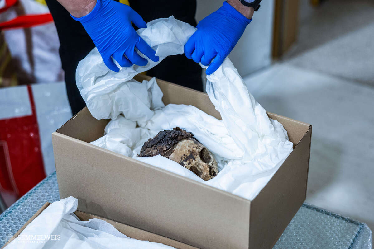
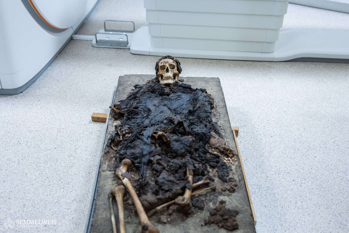
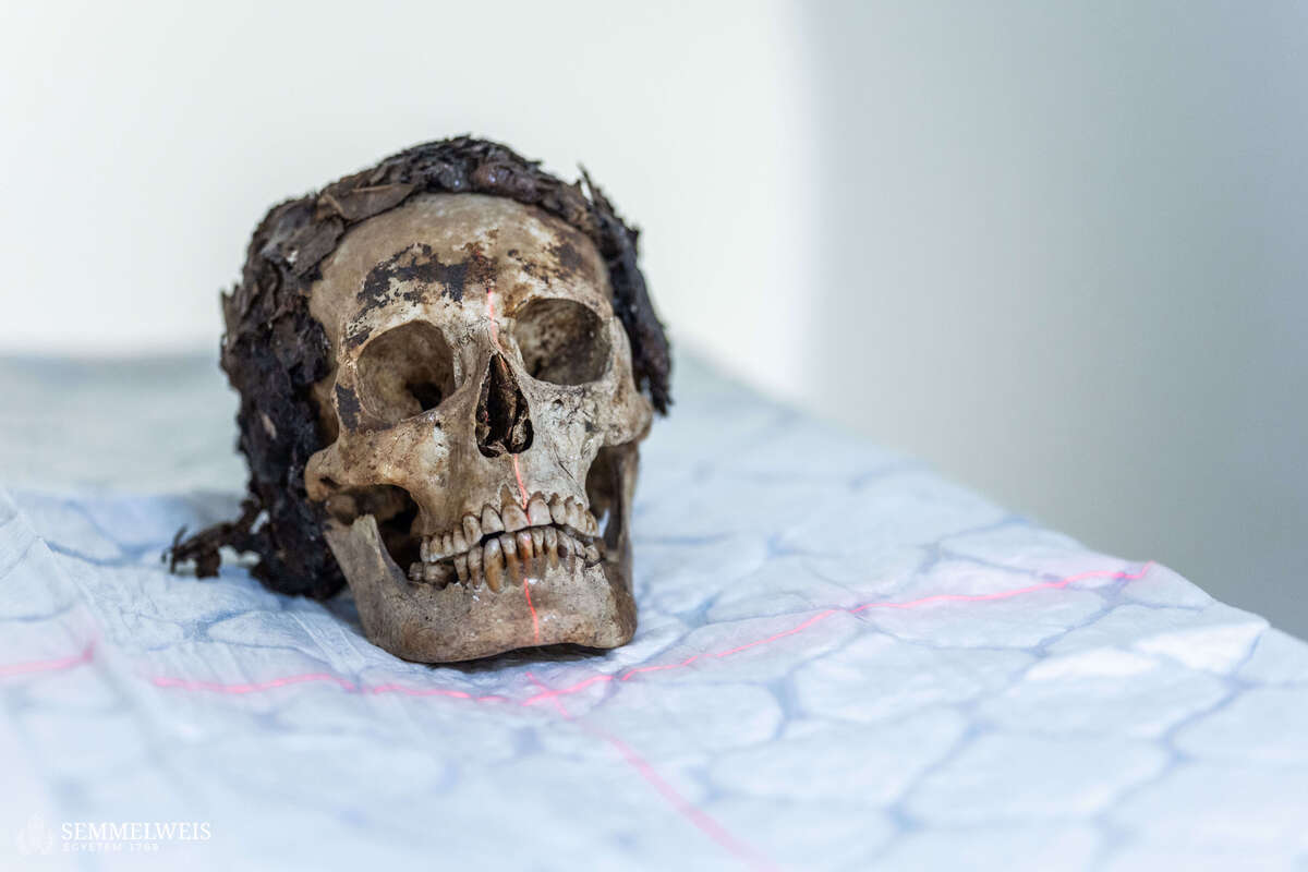
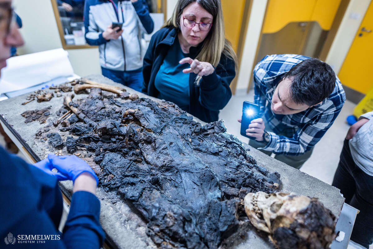
The mummy was found by chance during the construction of a detached house in the 3rd district of Budapest, on Jablonka Road, in 1962. The young woman whose body was discovered was between 20 and 25 years old, of fairly wealthy origin, probably lived in a villa in the neighborhood, and, in accordance with customs in the fourth century AD, was buried in the Roman villa estate’s own cemetery. The condition of the body, which had been preserved in some way, began to deteriorate rapidly after the sarcophagus was opened, so the archaeologists conducting the excavation tried to preserve the find as best they could with the means available at the time (glass cover, preservative material).
“After that, however, they were only able to carry out traditional archeological and anthropological examinations, which allowed them to determine her gender and approximate age, and the grave goods, i.e. the objects placed next to the deceased, were analyzed to identify the period in which she may have lived. Since no epitaph was found on the stone sarcophagus, and because it was common practice at the time to reuse them, the identity of the deceased and the cause of death remain unknown. It has not been examined with more modern tools and procedures ever since,” explained Dr. Orsolya Láng, archeologist at the BHM Aquincum Museum. Thus, it has also remained a mystery so far how exactly the body was preserved, whether any internal organs have survived, or whether other objects were placed under the burial shroud and funerary bandages.
Cutting-edge technology at the service of research
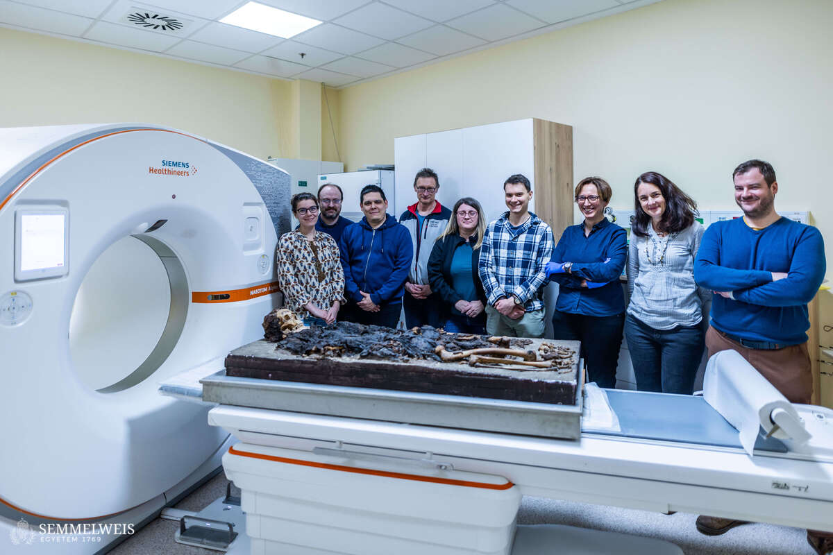
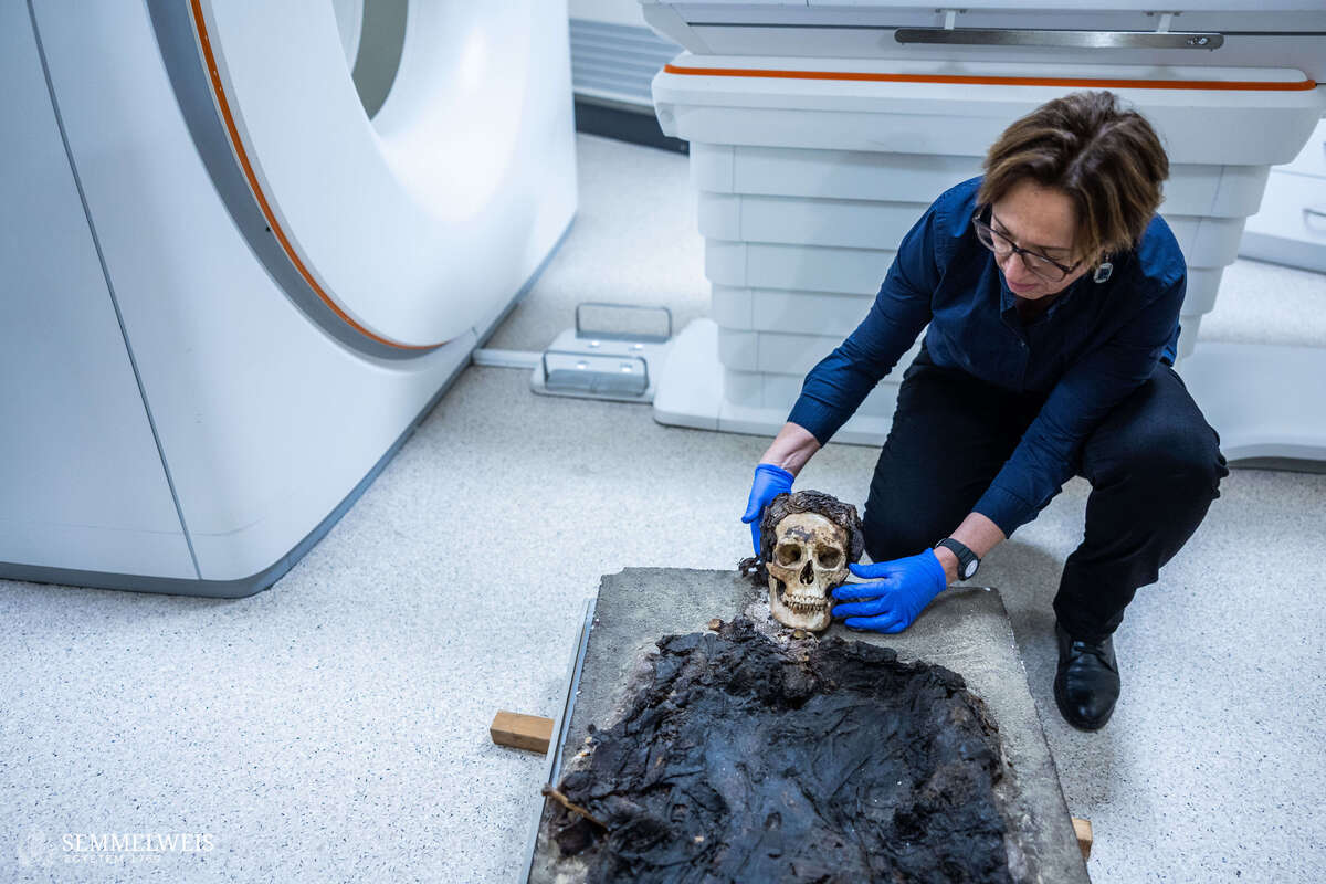
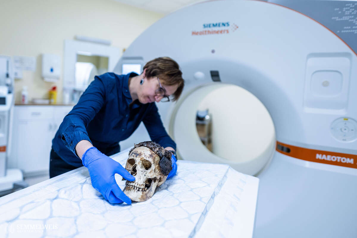
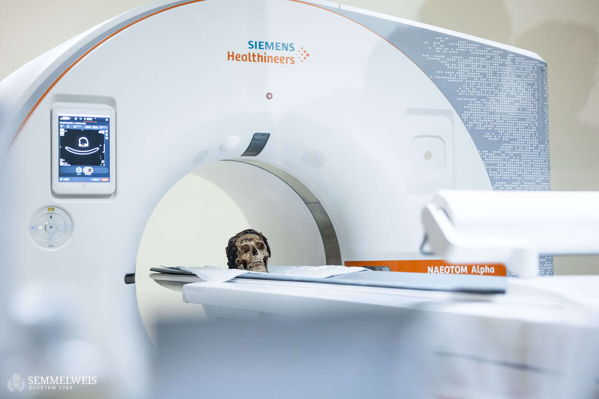
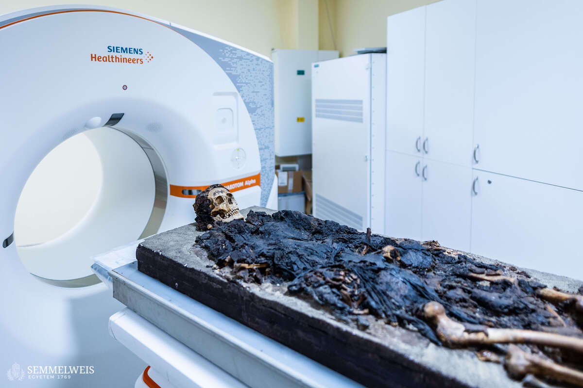
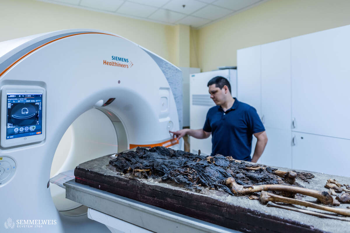
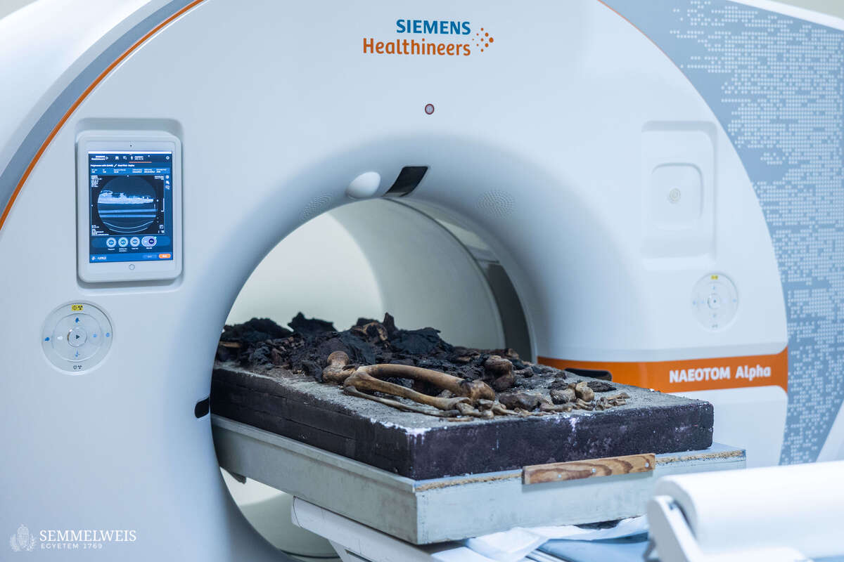
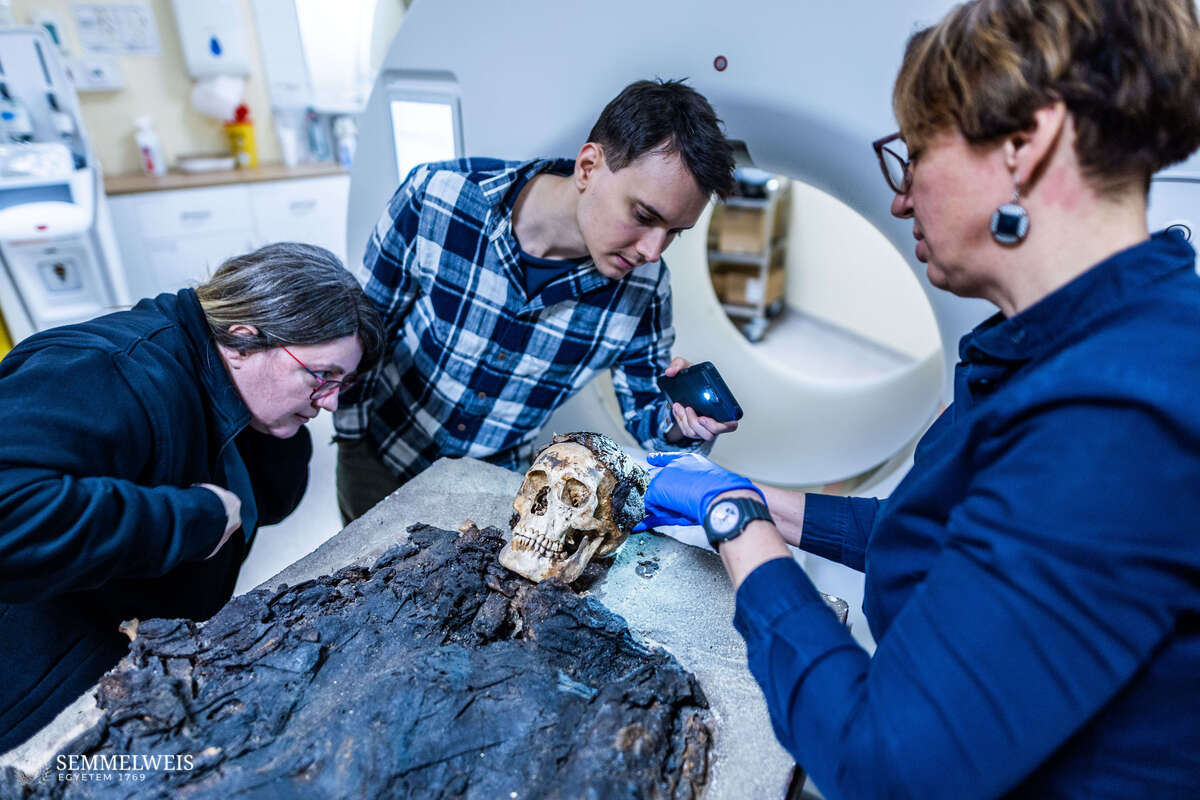
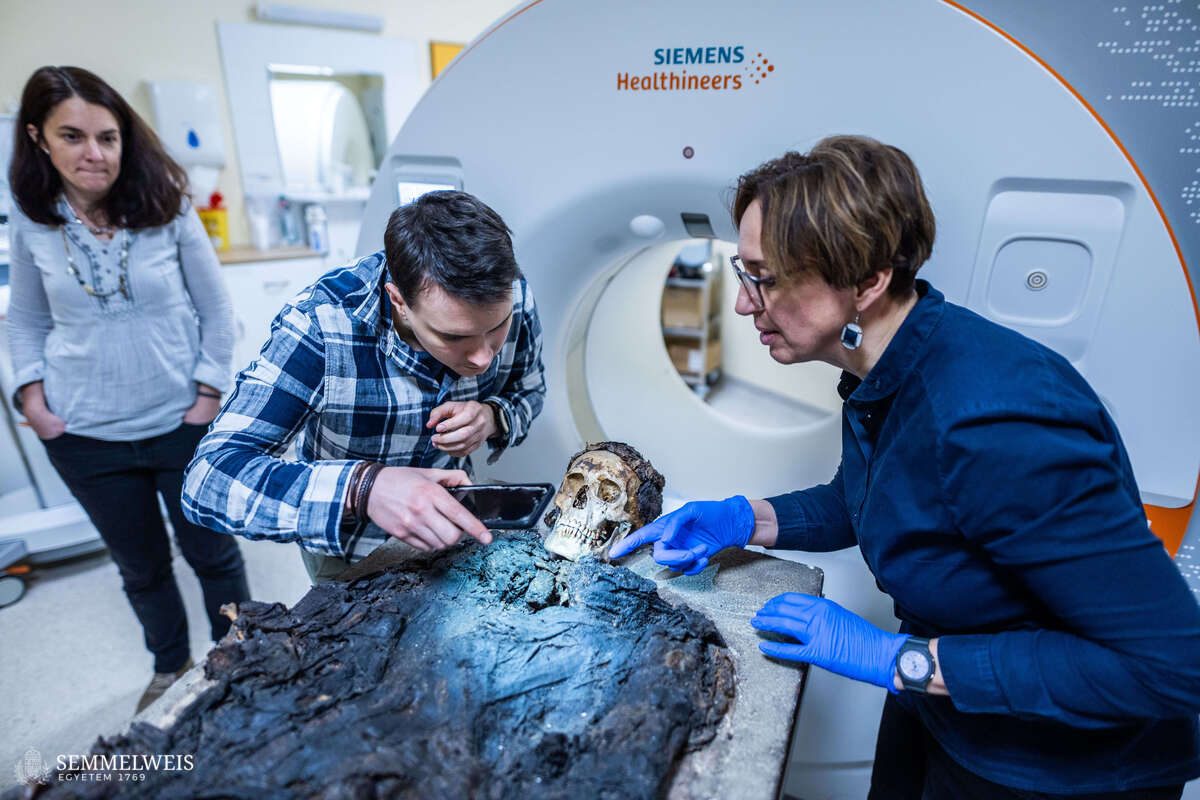
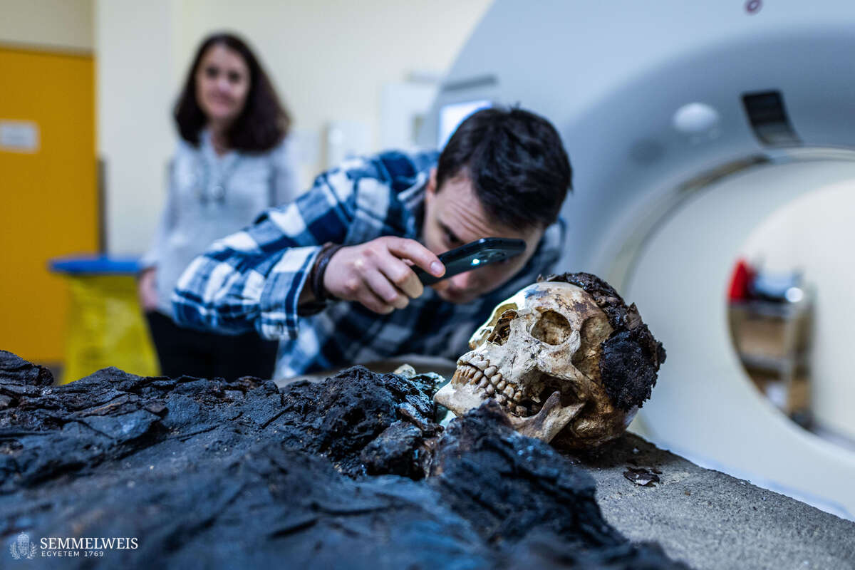
The mummy arrived at the Medical Imaging Center in the evening, outside of patient care hours, and was subjected to a computer tomography (CT) scan in accordance with hygiene regulations. The university has already put its third photon-counting detector CT scanner into operation – the first of these was put into service at OKK at the end of 2021, the second in OKK’s Emergency Radiology Unit, and the third in the Városmajor Heart and Vascular Center. All three serve research purposes in addition to patient care. Not only is medical and health science research being carried out, but the use of the devices for as many purposes as possible is also of interest to radiology and medical device development.
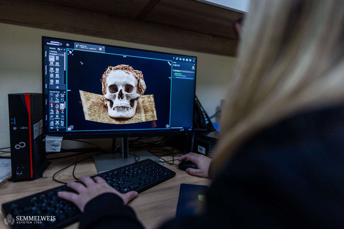
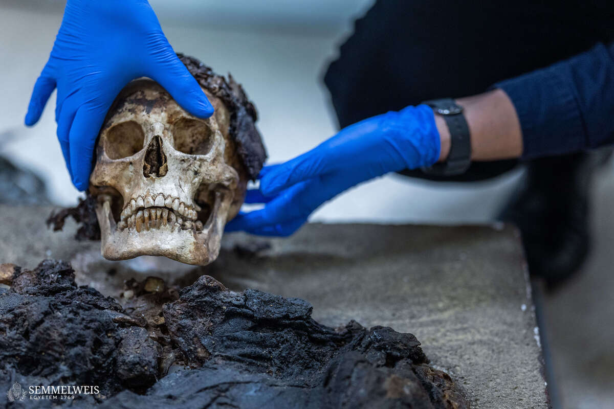
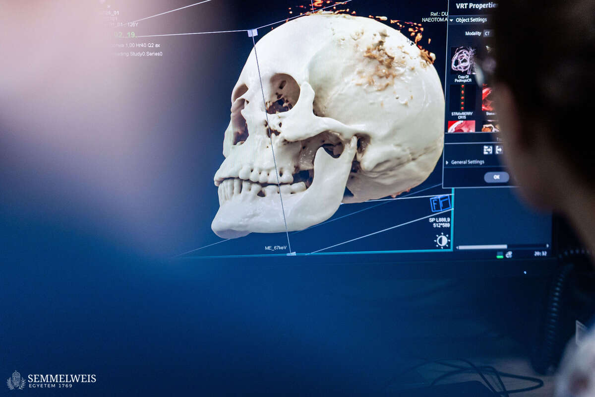
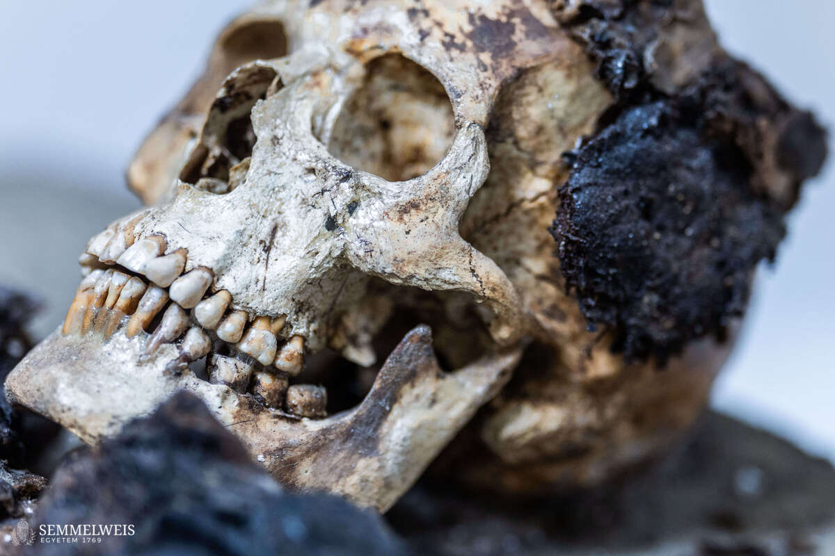
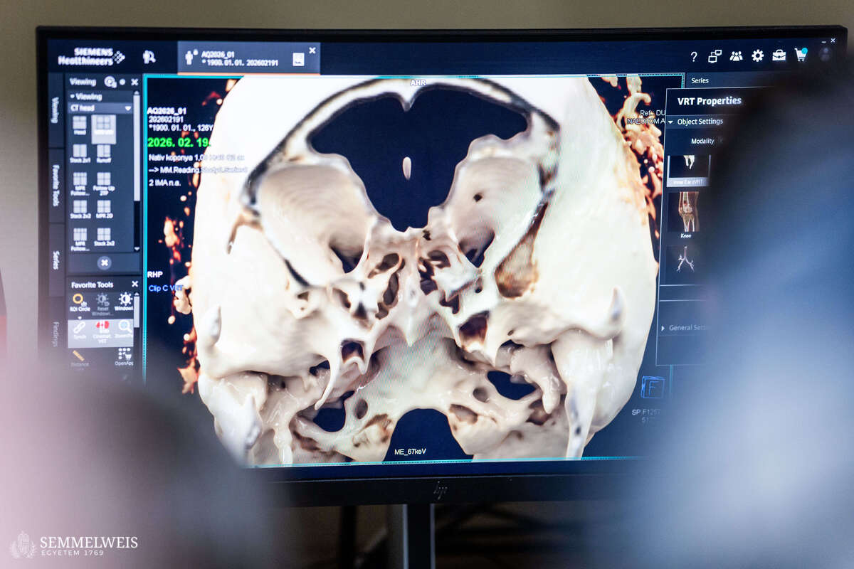
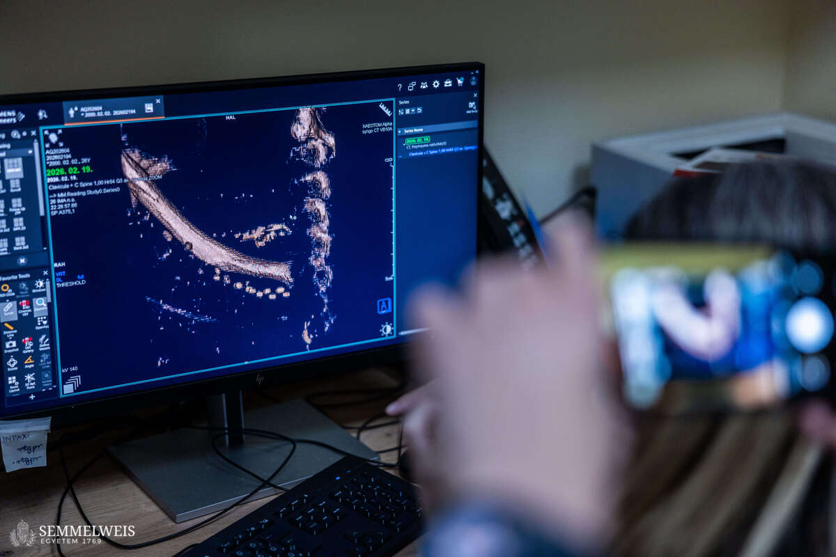
“Due to the specific nature of the find, separate images were taken of the skull after the auditory ossicles of one ear had been removed for subsequent DNA testing. First, the skull and the remaining human and other tissues were scanned with a fairly small slice thickness of 0.6 mm – revealing, for example, a minimal amount of sedimentary material in the cranial cavity, which will require further examination for a more accurate assessment. Then, for the planned facial reconstruction and further paleoanthropological and other examinations, we also took higher-resolution images with a slice thickness of 0.2 mm,” explained Dr. Ibolyka Dudás, who heads OKK’s post-mortem imaging working group.
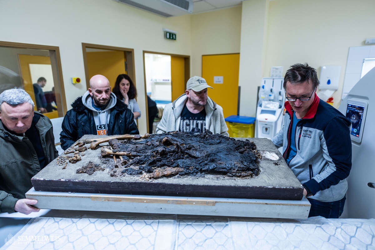
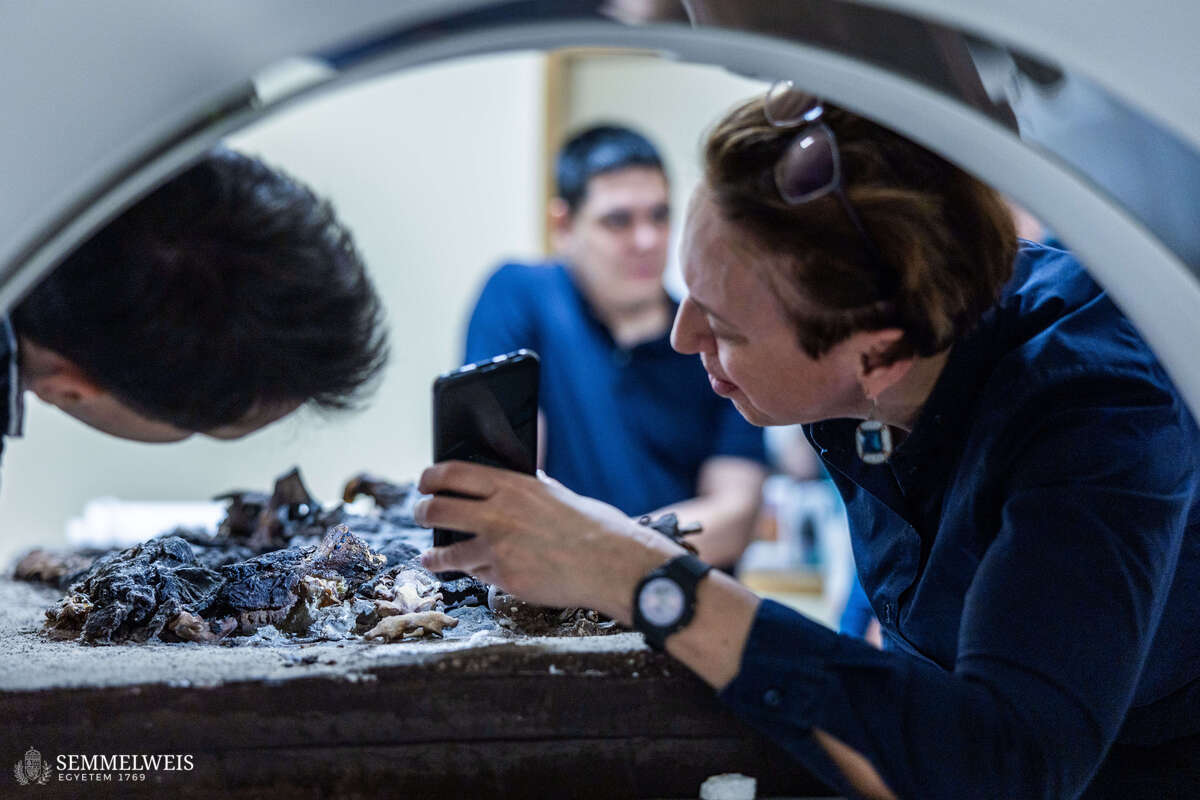
This was followed by an examination of the body, for which they managed to place into the machine not only the Styrofoam sheet under the body, but also the wooden frame supporting it. Semmelweis University has collaborated with archaeologists before – for example, with the Museum of Fine Arts, Budapest, during the CT examination of the Egyptian mummies in their collection, and it also played a role in the examination of the mummies found in Vác.
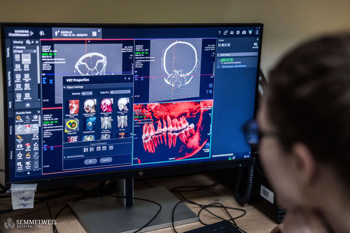
The radiologists then returned to the body parts that appeared most interesting based on the raw images and re-examined them at a higher resolution. This revealed that some decorative elements (beads) of the deceased’s clothing had survived under the burial shroud on certain parts of the body. The evaluation of the raw data from the images taken, the image reconstruction according to post-mortem examination protocols, and software processing will take place in the coming months.
Gallery
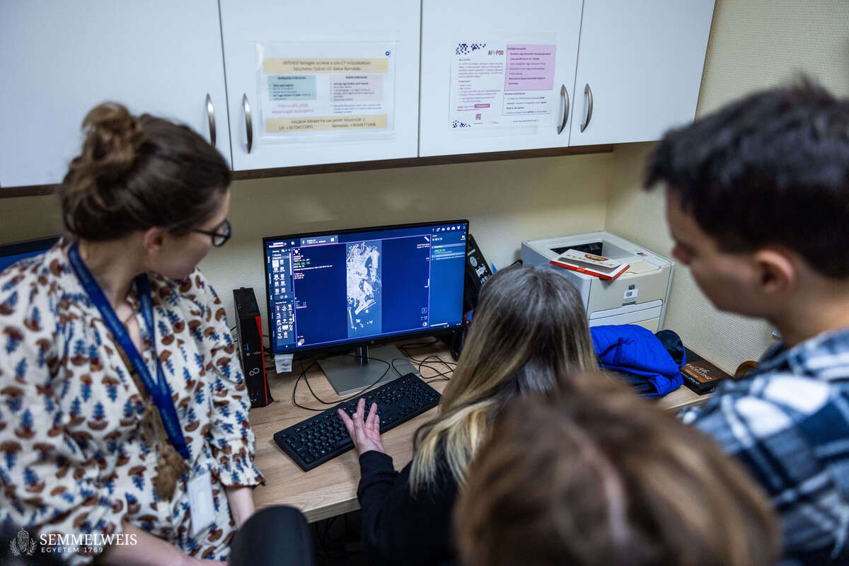
“Part of what we expect the CT scan to provide is a more detailed map of the body, including whether there are any changes, developmental abnormalities, or other diseases visible on the bones, or even signs of trauma, which could give us clues about the person’s life and possibly reveal the cause of death,” Anna Szécsényi-Nagy, Head of the research program and of the Institute of Archaeogenomics at the ELTE Research Centre for the Humanities, told our website. In further phases of the research, DNA testing will be performed on the extracted ear bone sample to determine the color of the deceased’s eyes, skin, and hair, as well as to find out where she came from, what other population she may have been close to, and whether she has any relatives in the ever-expanding Roman-era database.
In addition, the researchers want to use radiocarbon (C-14) dating to pinpoint the era in which she lived. They also want to shed light on her dietary habits and opportunities by examining stable isotopes of carbon and nitrogen, among others. A textile analysis will also be carried out to gain a more thorough understanding of the deceased’s clothing, the resin used for preservation, the bandages dipped in a salt solution, and the burial shroud. After processing the data from the images taken now and further examinations, the researchers would like to use the images made with the photon-counting detector CT in exhibitions and publications presenting the results, and they would also like to show the reconstructions to the general public.
Melinda Katalin Kiss
Translation: Dr. Balázs Csizmadia
Photos by Bálint Barta – Semmelweis University