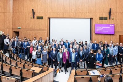
With approximately one hundred participants, the symposium held in the Semmelweis Salon focused on innovative fetal post-mortem pathology, exploring the role of minimally invasive autopsy and fetal imaging in diagnostics. Dr. Owen Arthurs, Professor of Radiology at Great Ormond Street Hospital in London, and Dr. Ciaran Hutchinson, Consultant Pediatric Pathologist, gave richly illustrated presentations on how state-of-the-art imaging techniques (CT, micro-CT, MRI) have been integrated into everyday pathological diagnostics and how they facilitate faster, targeted diagnostics and tissue-level, pinpoint morphological data collection (deep phenotyping) based on thousands of cases in their database.
Dr. Pál Maurovich Horvat, Director of OKK, and Dr. Ibolyka Dudás, Chief Physician of OKK’s Department of Radiology, presented their experiences gained at Semmelweis University, while Dr. Artúr Beke, Associate Professor at the Department of Obstetrics and Gynecology, spoke about the genetic aspects of fetal diagnostics. Dr. Béla Molnár, Research Professor at the Department of Internal Medicine and Oncology, founder and owner of the startup 3DHISTECH, reported that his long-held dream of 3D digital tissue-level imaging has come true with the company’s latest development, the micro-CT scanner. Micro-CT imaging is ideal for postmortem fetal diagnostics, as demonstrated by PhD student Lili Száraz and Dr. Owen Arthurs.

During the afternoon workshop, Dr. Attila Fintha and Dr. Noémi Jákob, members of the PKRI perinatal working group, as well as Dr. Ibolyka Dudás and Dr. Owen Arthurs presented illustrative cases using spectacular and innovative imaging techniques (photon-counting CT, CT, micro-CT).
The program is accredited by the London-based Royal College of Pathologists, which has more than 13,000 members worldwide. The organizers, Dr. Beáta Hargitai and Dr. Tamás Marton, Associate Professors at the Department of Obstetrics and Gynecology, emphasized that the approximately 100 participants included specialists from numerous university centers in Hungary and abroad, representing the fields of obstetrics, radiology, fetal medicine, and pathology. Attendees came from various cities, including London, Edinburgh, Birmingham, Liverpool, Paris, Athens, Utrecht, Debrecen, and Pécs.
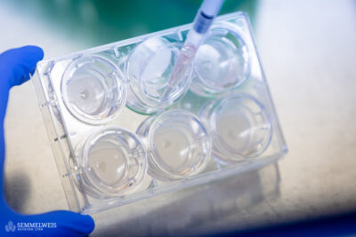
Non-invasive postmortem examination techniques make it possible to perform autopsies with minimal incision out of respect for the deceased, using various imaging techniques such as MRI, CT, micro-CT, or postmortem ultrasound, pointed out Dr. Tamás Marton. These methods can be used to examine the anatomy of the fetus and identify developmental abnormalities, primarily in relation to the central nervous system; they also enable targeted tissue sampling in a minimally invasive manner. The procedure requires a multidisciplinary approach involving radiologists, pathologists, and fetal medicine specialists. Non-invasive and minimally invasive autopsies are a progressive trend that could replace traditional autopsies, he added.
Group photo and text by Dr. Beáta Hargitai, Dr. Tamás Marton – Department of Obstetrics and Gynecology
Cover and illustration: 3D reconstructed image of a seven-week-old fetus using micro-CT scanning; image credit: London working group, Shelmerdine et al., Prenatal Diagnosis 2018
Translation: Judit Szabados-Dőtsch