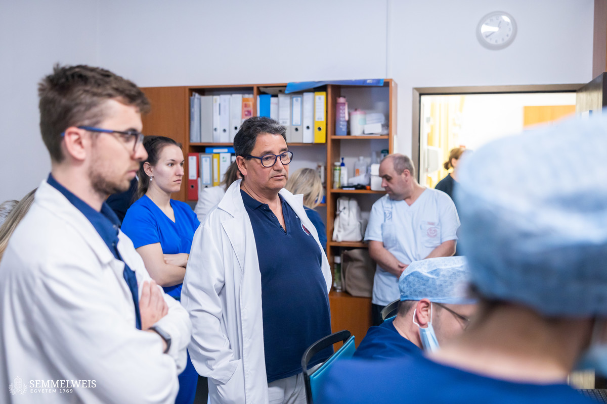
 “I have had the opportunity to track the development of catheter ablation throughout my career: in the mid-1990s, I was the first to introduce catheter ablation at the Városmajor Heart and Vascular Center, and we were pioneers in Hungary in performing ablation of atrial flutter, one of the most common cardiac arrhythmias. And now, we have been the first in Hungary to perform ablation of atrial flutter with the help of an MRI machine,” said Dr. Béla Merkely, Rector of Semmelweis University and Director of the center. As he pointed out, the Research Laboratory of the center in Városmajor has performed several ablation tests and developments of ablation catheters. The introduction of MRI-assisted procedures had been planned for more than five years before the first surgery was performed.
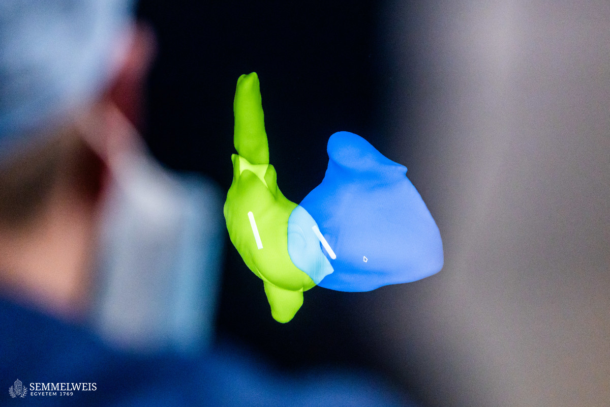
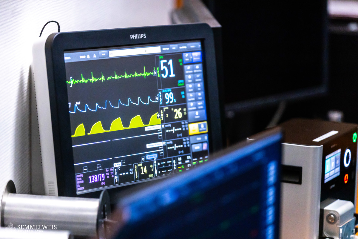
One of the difficulties of catheter ablation is that we usually navigate in the heart by using electrical signals and X-rays. However, the X-ray does not give us any feedback on the effect of the different ablation energies on the heart, or the amount of lesion, i.e., targeted damage. A new option to solve this challenge is to perform ablation surgery in conjunction with magnetic resonance imaging, as MRI scans the heart at the tissue level and at the same time can create an anatomical map of the organ to help navigation. – Dr. Béla Merkely
As the rector explained, the intervention is currently used for certain disease groups in Hungary, but with the advancement of technology, it will be possible to use it in more and more areas. In the future, even catheter interventions for stroke may be performed with MRI.
Ablation is the term used to describe interventions that block faulty heart signals. In essence, tiny scars are created in the areas of the heart muscle responsible for the heart rhythm irregularity, preventing it from generating or transmitting faulty heart signals. Ablation can be carried out by using various techniques, such as heat or cold, or other energy-based methods. It is a minimally invasive procedure with fewer complications and faster recovery compared to traditional surgical options.
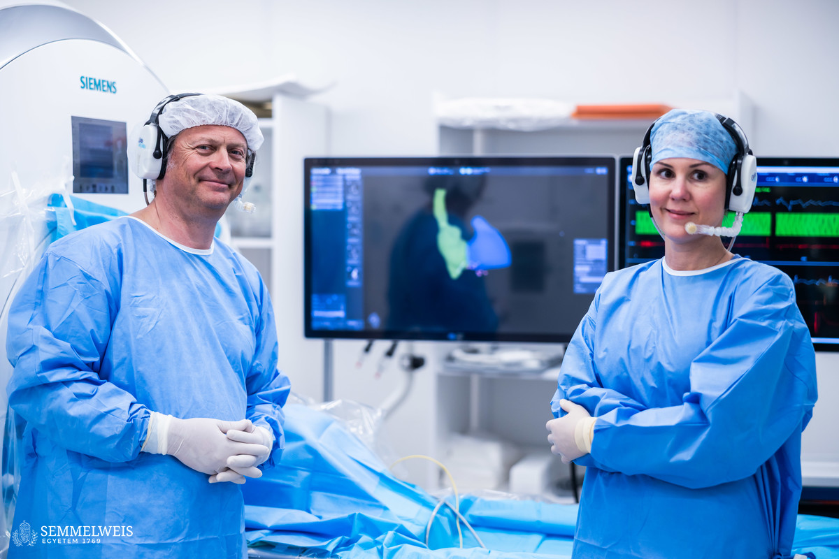
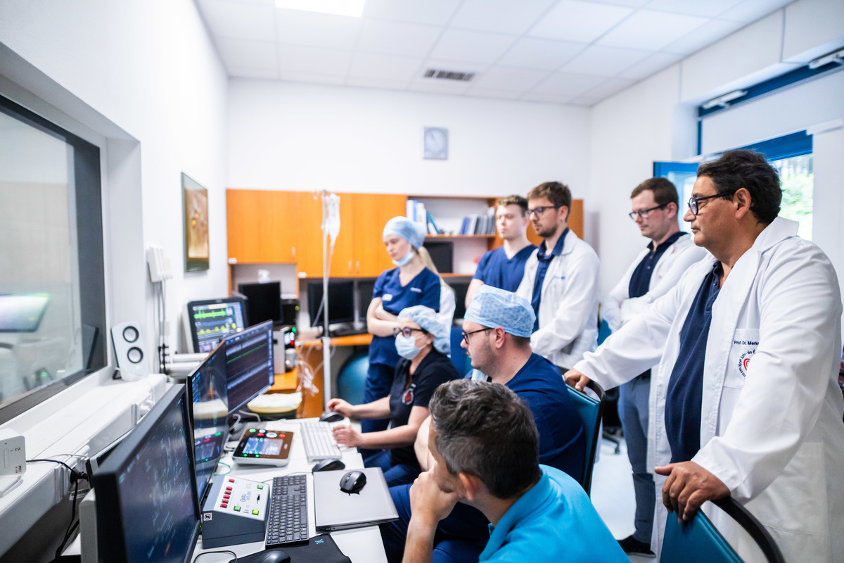
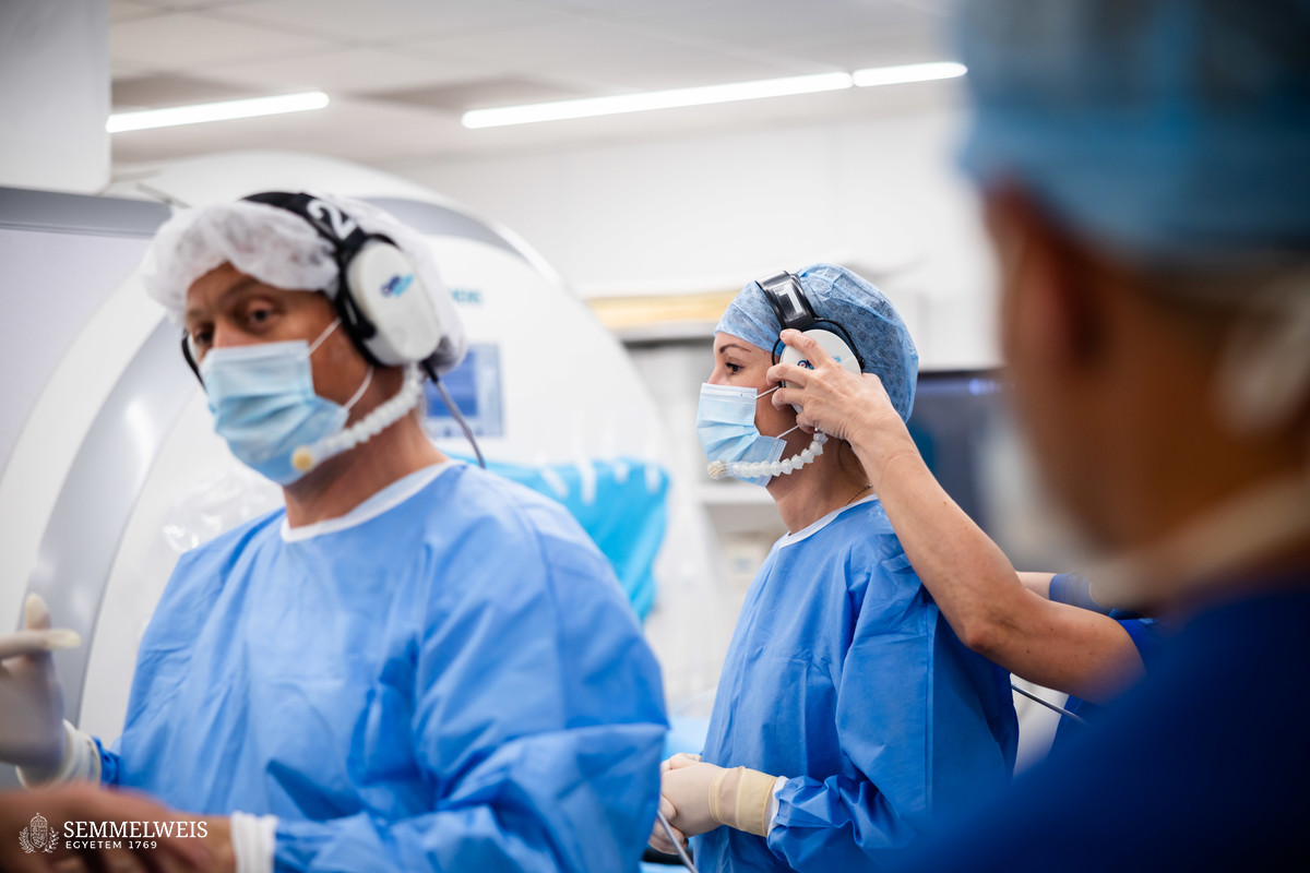
The intervention was performed by Dr. László Gellér, Head of the electrophysiological working group at the Department of Cardiology, and Dr. Klaudia Vivien Nagy, Associate Professor at the Department of Cardiology and the Department of Aviation and Space Medicine. As Dr. László Gellér pointed out, one of the biggest advantages of MR-ablation was that, unlike conventional X-ray interventions, the patient was not exposed to radiation, and MR was the best at revealing the substrates responsible for cardiac arrhythmias, i.e., scars and foci. This is why the intervention can make the treatment of arrhythmias even more effective than before. “We have high expectations for this technology, which may even make the treatment of ventricular tachycardia possible in the future,” he added.
Gallery
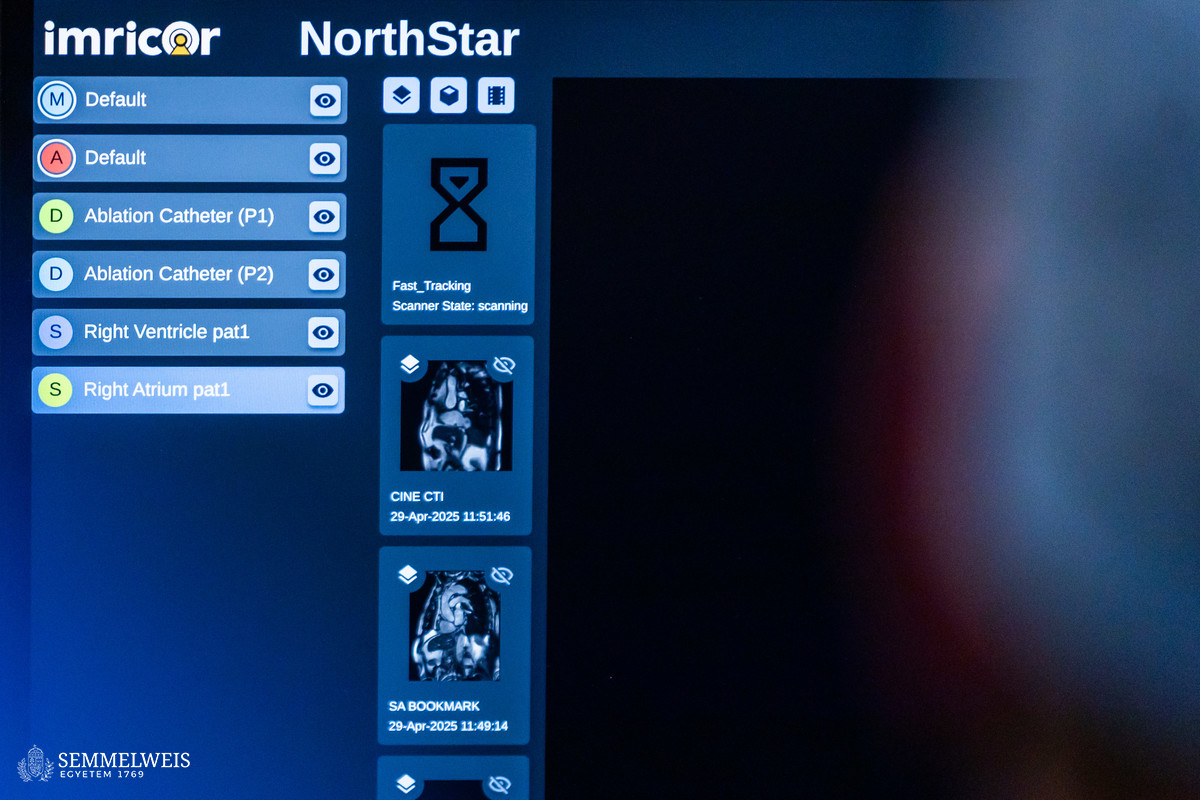
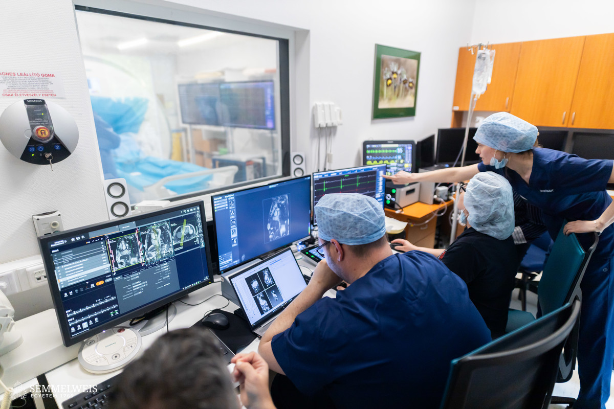
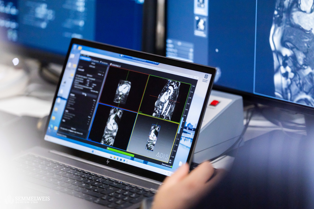
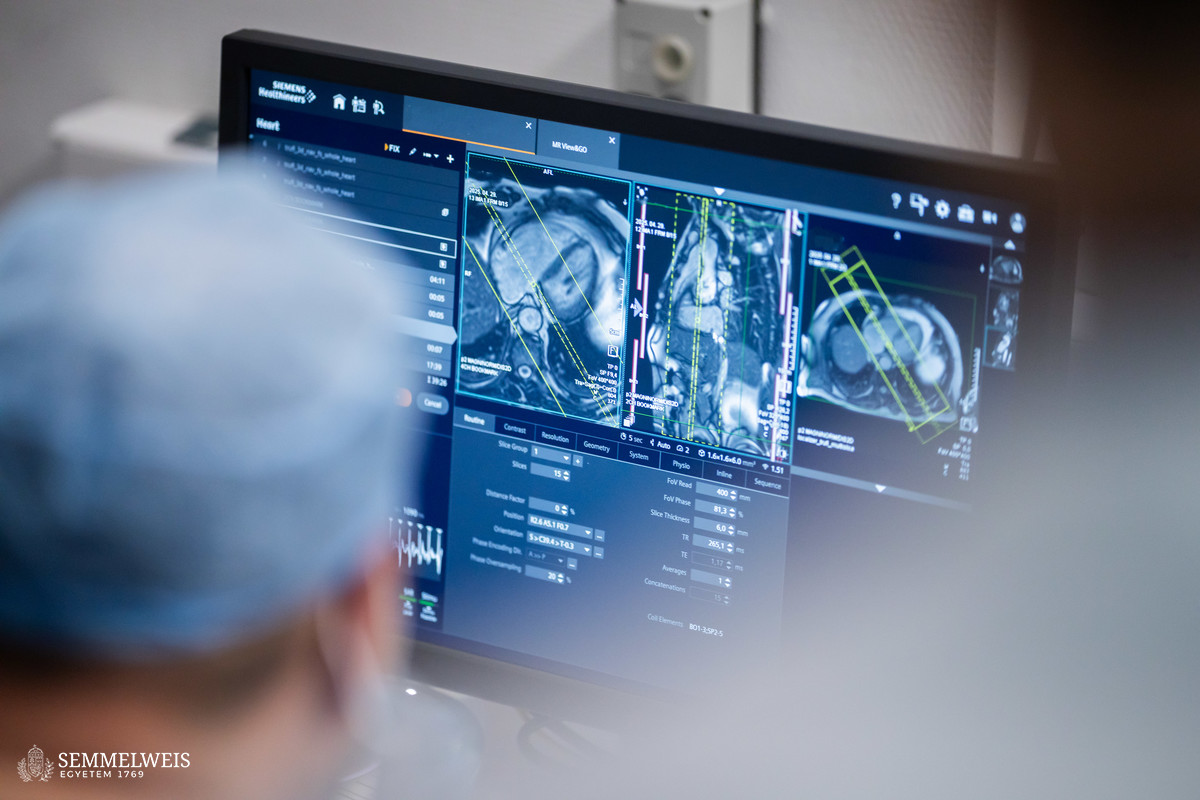
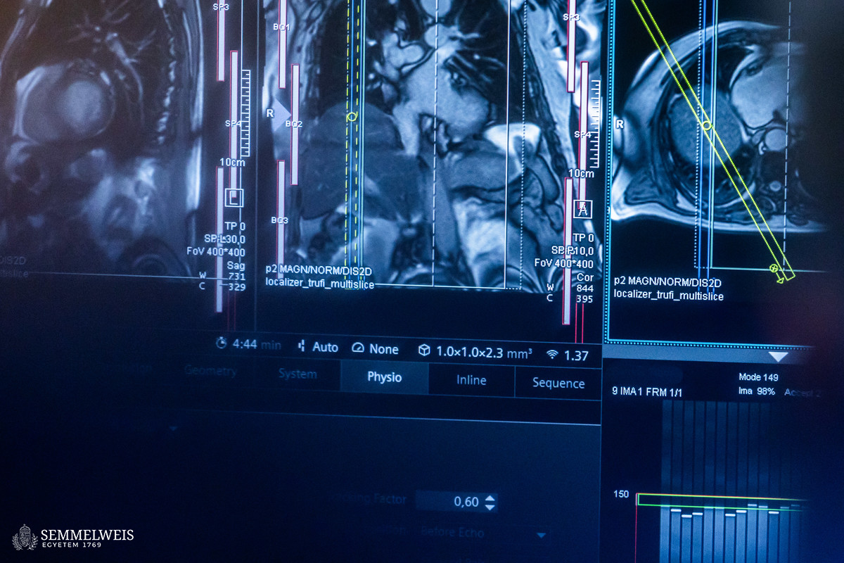
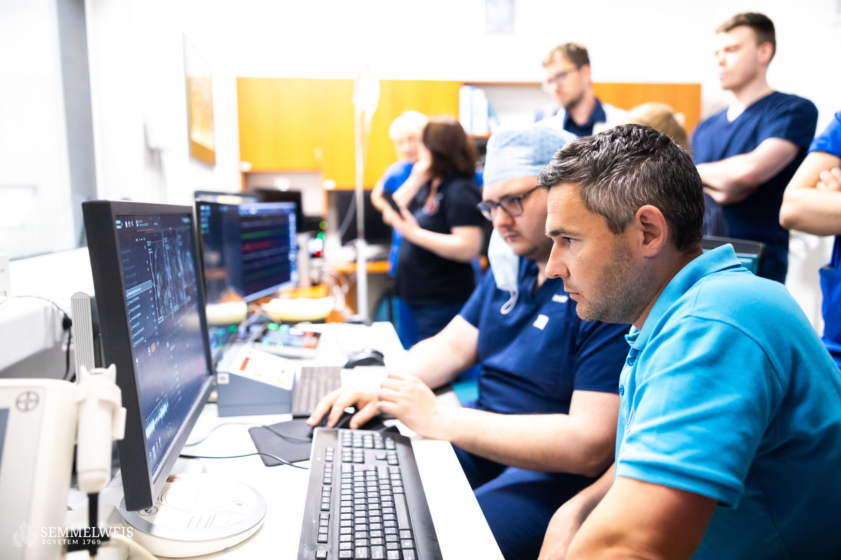
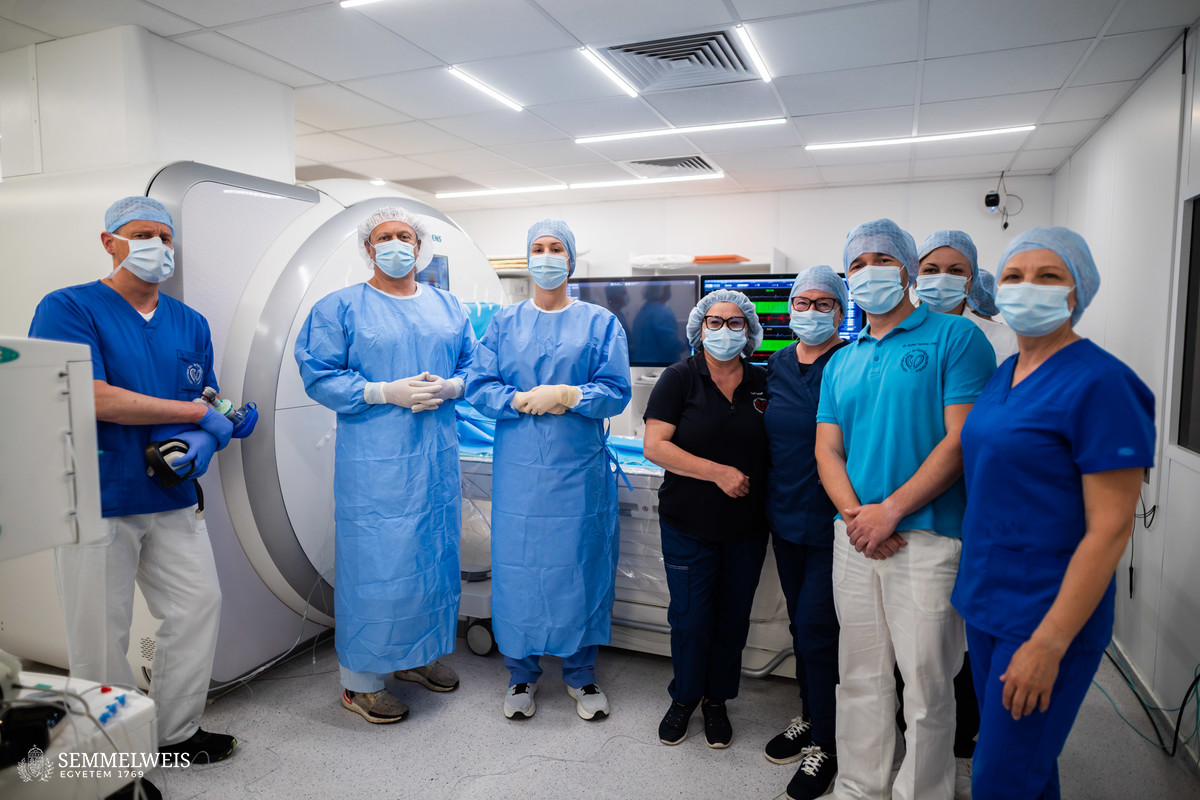
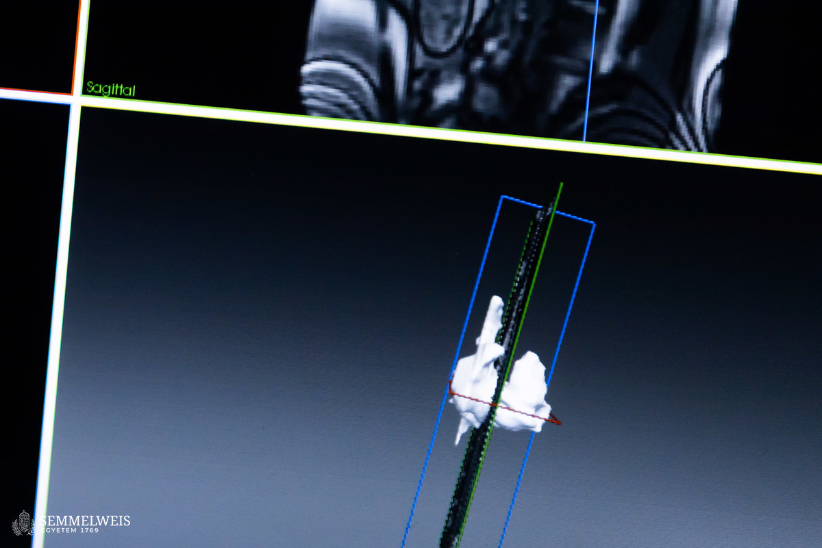
Dr. Klaudia Vivien Nagy emphasized that MRI provided an incomparably more accurate picture of the heart than traditional two-dimensional X-rays. However, MRI requires a special environment: no metal or magnetizable devices can be brought into the examination room – hence the need to develop special MRI-compatible catheters. The intervention used equipment from the US company Imricor, and their new mapping software was used for the first time in the region at the Városmajor Heart and Vascular Center.
Ádám Szabó
Translation: Judit Szabados-Dőtsch
Photos by Bálint Barta – Semmelweis University