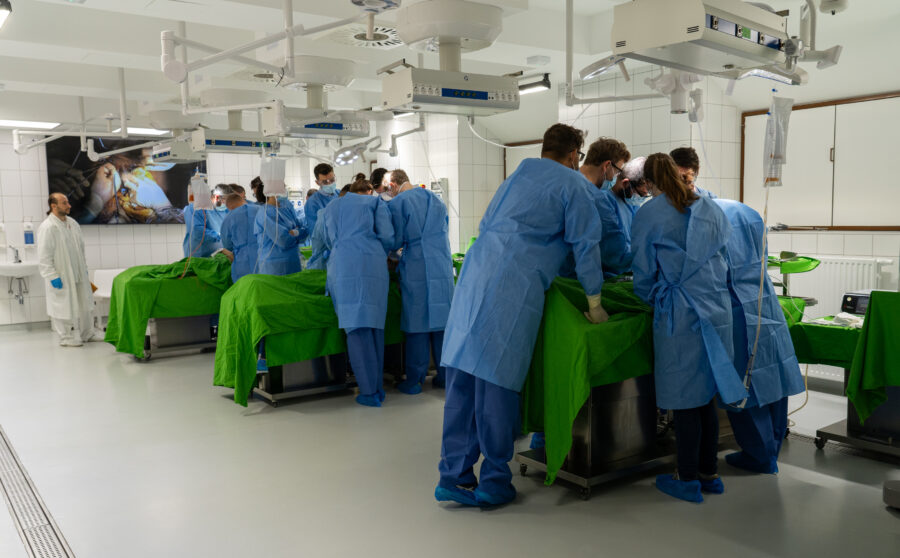
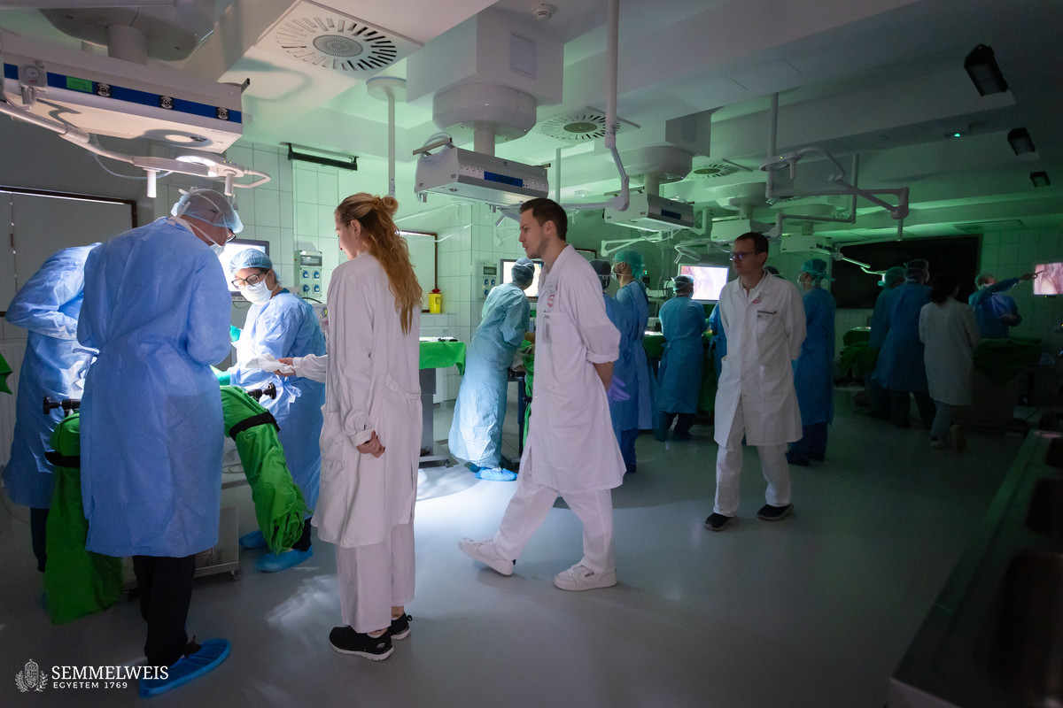
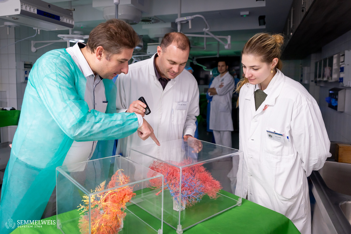
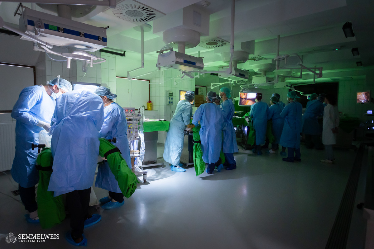
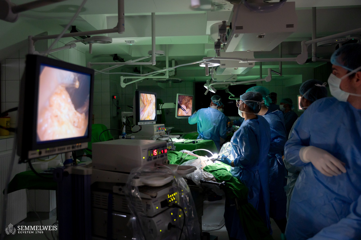
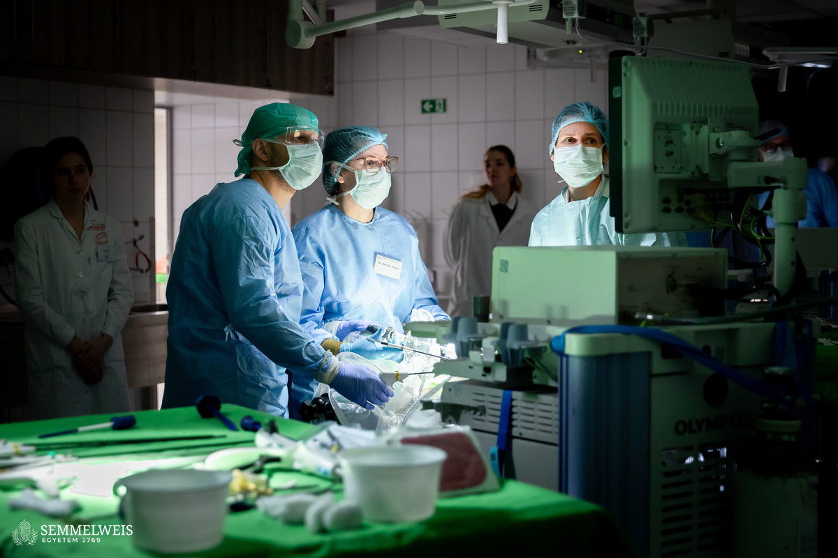
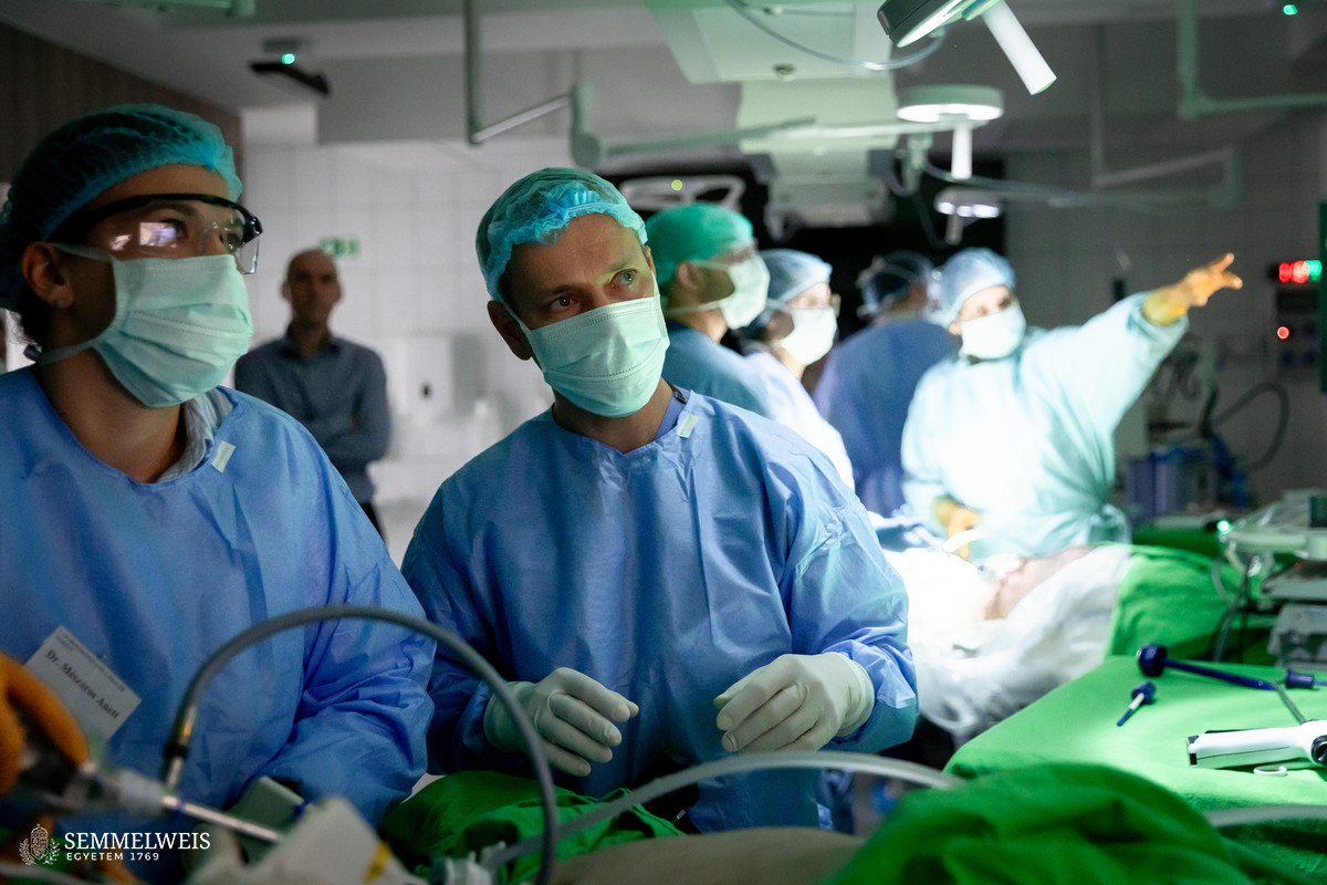
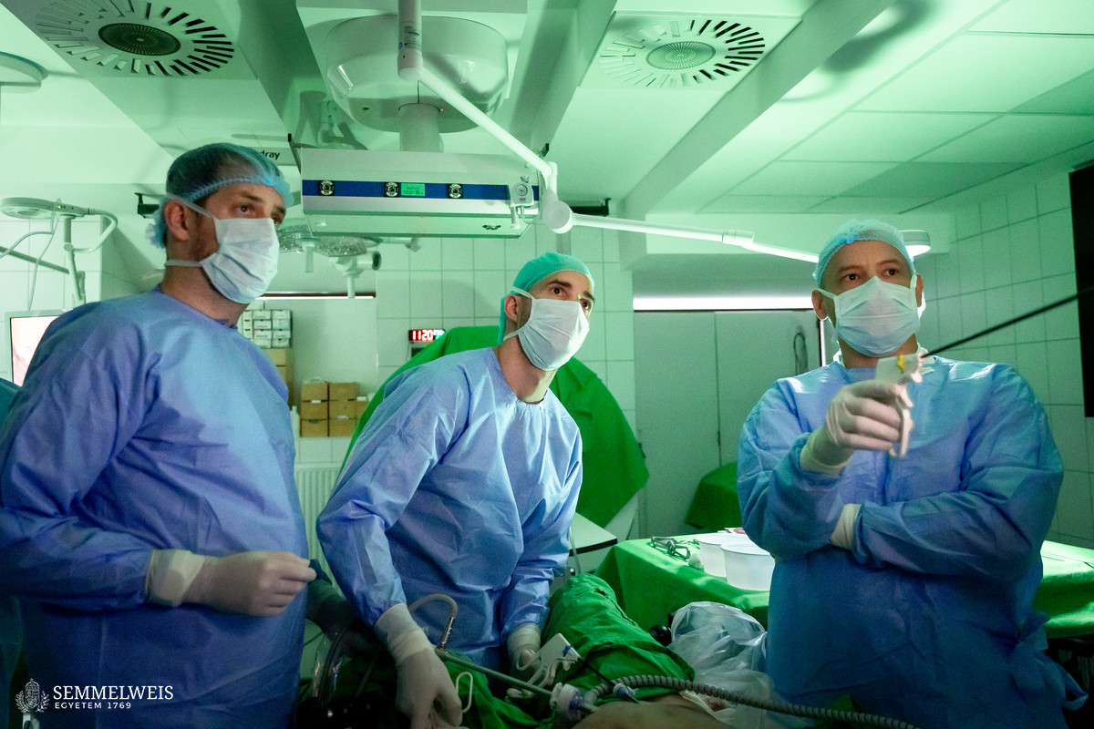
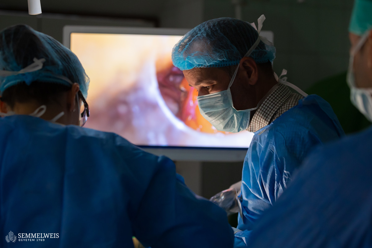
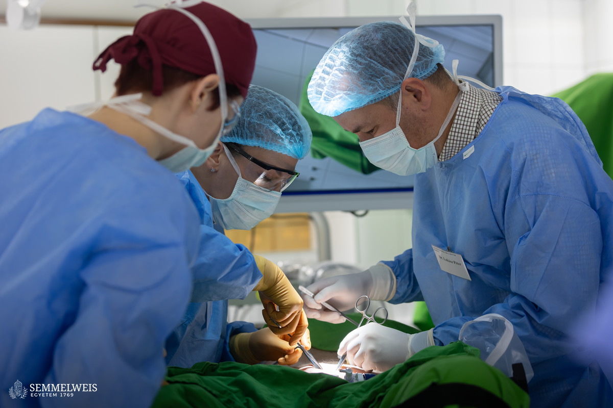
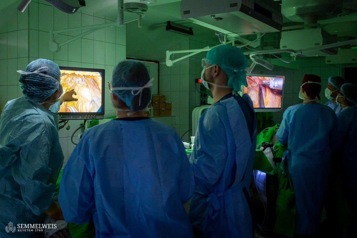
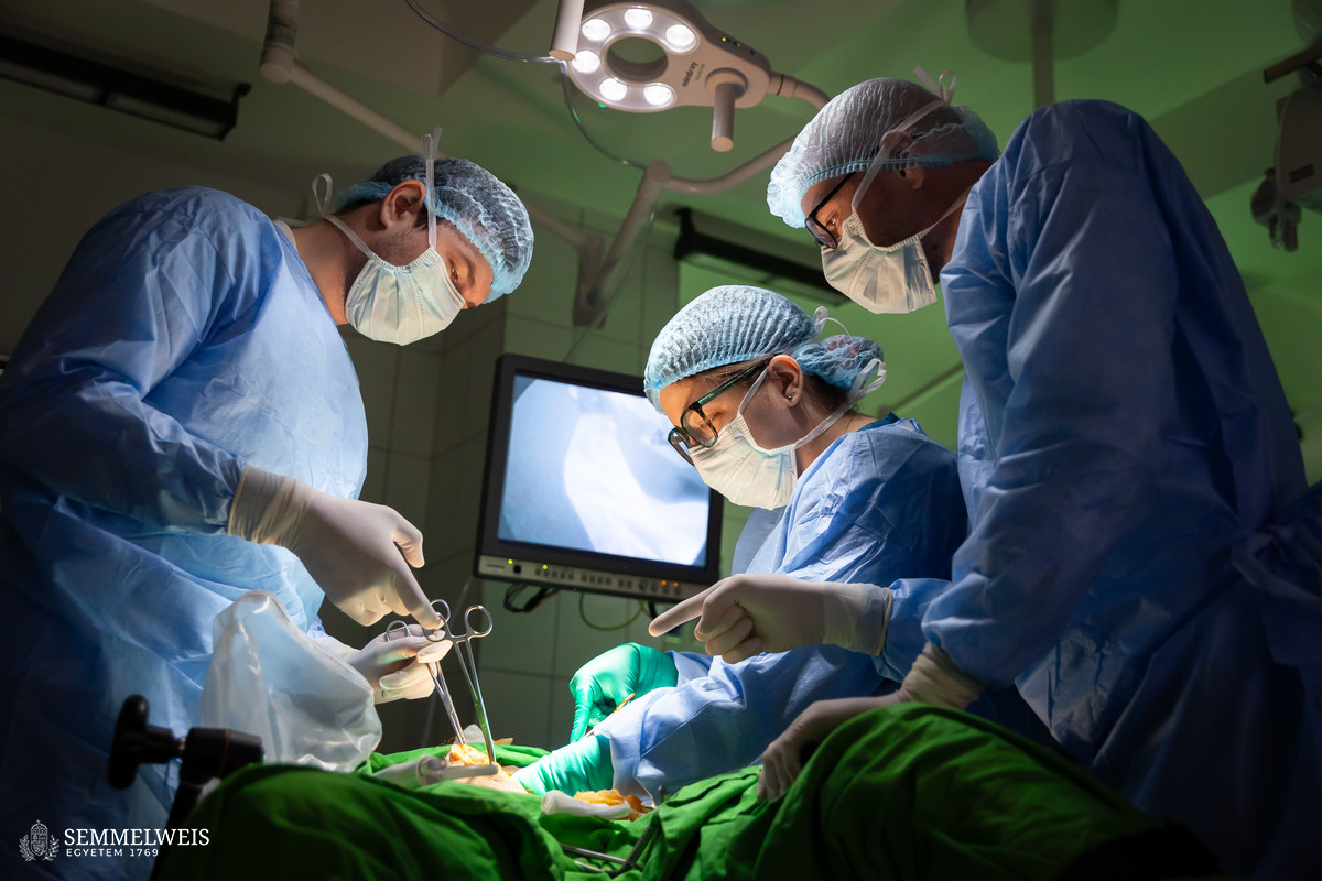
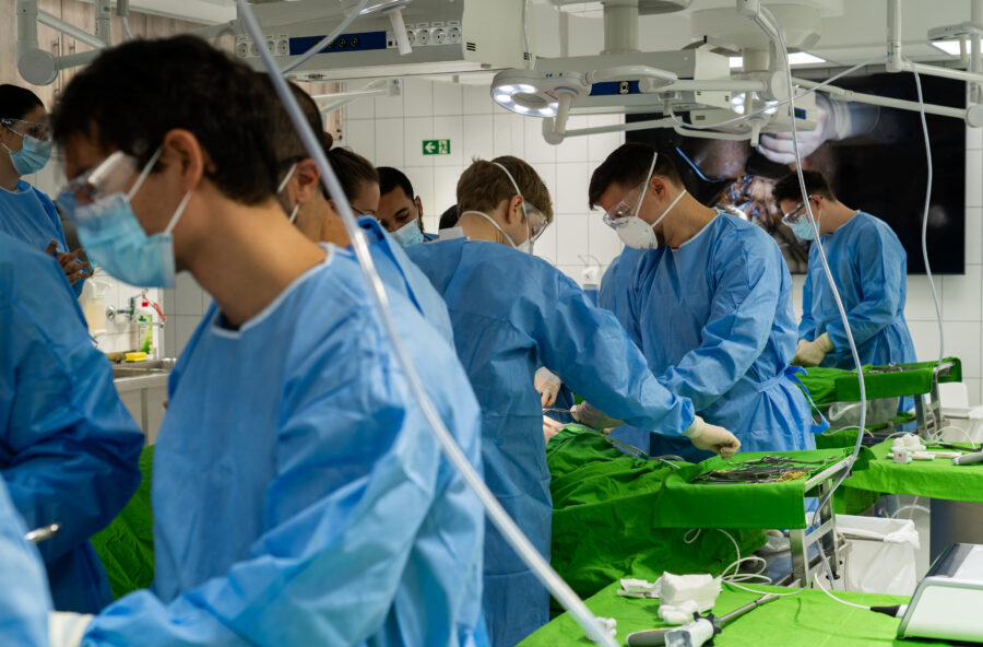
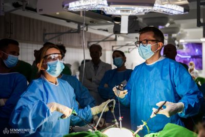
There are few cadaveric medical vocational and specialized training courses across Europe, yet these hands-on training sessions are an undeniably effective way of developing surgical manual skills. Semmelweis University’s courses offer a unique opportunity among surgical training courses due to the use of a special tissue fixation technology developed by Viktor Pankovics, an embalmer at the Department of Anatomy, Histology and Embryology. This type of preparation, which results in soft tissues, vascular formations, and organ complexes very similar to their counterparts in the operating room (even bleeding can be simulated), provides surgeons with a real operating room situation and a unique opportunity to practice. In addition, the empirical extension of the three-dimensional clinical anatomical approach is also a key part of the courses. The organs and the different vascular variations were demonstrated on vascular casts from the invaluable collection maintained by Dr. Ágnes Nemeskéri and Dr. Mátyás Kiss, as well as on anatomical preparations made by students from the Students’ Scientific Association. During the two sessions of the course series, surgeons were able to practice and memorize the steps required to perform pancreatic surgery using open surgical and minimally invasive laparoscopic techniques, and they also had the opportunity to carry out operations on the liver and colon, as Dr. Tamás Ruttkay, Assistant Professor at the Department of Anatomy, Histology and Embryology, told our website.
The cadaveric abdominal surgery course series started with open, hands-on pancreatic surgery training. As Dr. Ákos Szücs, Associate Professor at the Department of Surgery, Transplantation and Gastroenterology (STéG), and professional leader of the training – in addition to Dr. Tamás Marjai, Specialist Physician at STéG – noted, pancreatic surgery is a complex procedure, and there are few opportunities for young specialists to practice safely. They are often able to perform sub-tasks only at the operating table, and this was the first time they had the opportunity to perform a full operation, from skin incision to skin suture, on the cadavers with artificial tumors.
The cadaver itself, thanks to the preparation technique, created a very lifelike environment, even allowing for bleeding to be simulated, Dr. Ákos Szücs stressed, adding that the saturation of the vascular formations and the treatment of the bleeding caused by the injury also evoked a realistic surgical environment, which is unique not only in Hungary but also internationally. The use of conventional fixation techniques is much less able to reproduce the texture of real living human tissue and does not provide the difficulties to be encountered in a real surgical environment, in contrast to the technique developed at the department.
During the two-day training, after the theoretical introduction, the participants were first trained on organ complexes to improve the surgical techniques of anastomosis after tumor removal, such as the reconstruction and connection of the alimentary canal, stomach, and bile ducts; they also had the opportunity to examine and treat cases related to blood supply on the prepared specimens. On the second day, they were able to perform or assist throughout the entire pancreas head removal surgery.
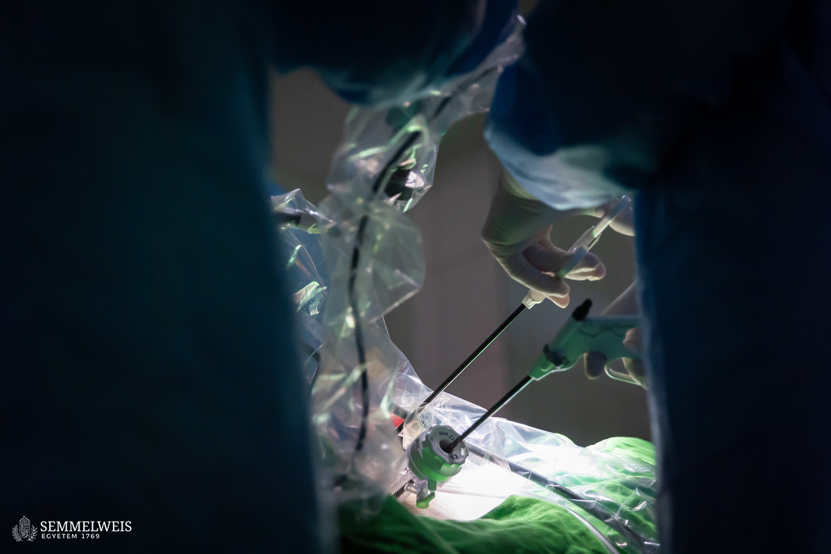
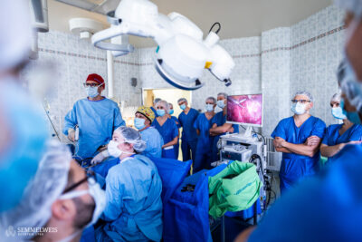
In the second session of the cadaveric pancreatic surgery course, also led by Dr. Tamás Marjai and Dr. Ákos Szücs, participants were taught the minimally invasive laparoscopic technique. The aim of this training was to practice the laparoscopic procedure, which is considered the gold standard for the removal of the pancreatic tail, most often necessitated by a tumor. For this operation as well, the unique fixation technique created a tissue environment for the removal of the pancreatic tail that was very similar to the real surgical situation, explained Dr. Ákos Szücs. The training was a great success, similarly to the open surgery version, and with the course planned for next year the organizers are already preparing for the international arena, as this type of training is considered to be filling a niche in Europe and could thus play a pioneering role in the training market, Dr. Ákos Szücs added.
One session of the course series was dedicated to liver surgery, entitled “Applied Liver Anatomy in Oncosurgery and Transplantation,” where hands-on cadaveric training was provided using specially preserved model preparations. When practicing liver surgery interventions, a lifelike, transparent tissue environment is particularly important due to the special anatomical environment of the organ, emphasized Dr. Attila Szijártó, Director of STéG and one of the professional leaders of the course. He said that the course focused on acquiring the routine of tumor surgery, but the participating surgeons, who had previously only been able to practice on silicone models, also had the opportunity to familiarize themselves with the steps of transplantation and practice and memorize the necessary techniques. Dr. Attila Szijártó considered the training to be intensive and very useful, which he said could not be replaced by any other technique and should be promoted in international forums.
The last session of the course series was devoted to the new radical colon surgical technique of complete mesocolon excision (CME), which is expected to bring about a significant improvement in patients’ life expectancy in the long term, pointed out course leader Dr. Balázs Bánky, Associate Professor at STéG. Colon tumors are operated on at the university using this technique, and the course offers a safe training opportunity for both surgeons from and outside the university, Dr. Balázs Bánky added. The safe professional environment and the high-quality tissue layers and structures of the cadavers, as well as the state-of-the-art set of laparoscopic tools all contributed to the positive feedback from the participants, he stressed.
Anita Szepesi
Translation: Dr. Balázs Csizmadia
Photos by Boglárka Zellei – Semmelweis University; Dorka Székely
