The staff of four clinics of Semmelweis University organized a unique cadaver course aimed at mastering the practice of gastroscopy and colonoscopy for gastroenterologist candidates at the beginning of their endoscopy training: the Department of Anatomy, Histology and Embryology (Dr. Tamás Ruttkay, Dr. Gábor Baksa), the Department of Internal Medicine and Oncology (Dr. László Barkai, Dr. Ákos Iliás), the Institute of Pancreatic Diseases (Dr. Bálint Erőss), and the Department of Surgery, Transplantation and Gastroenterology (Dr. István Hritz). Since the stomach and intestinal system quickly decompose after death, it was not possible to perform gastroscopy on a cadaver before. The unique training was based on a new tissue preservation technology developed by the department’s embalmer, Viktor Pankovics. Specialist doctor candidates from all over the country applied for the course announced and accredited by OFTEX, the Hungarian medical doctor training system, and the seven participants were guided by four gastroenterologists. The next training is planned for the end of May, said Dr. Tamás Ruttkay, assistant professor at the Department of Anatomy, Histology and Embryology.
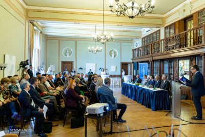
The one-day program entitled “Introductory gastroenterology endoscopic »hands-on« course on cadaver” began with a short theoretical presentation, during which the anatomists explained the special preparation technique that makes cadavers suitable for endoscopic examination, and then the participants were given a summary of the necessity and risks of endoscopic interventions by the gastroenterologists, outlined the details Dr. László Barkai, teaching assistant at the Department of Internal Medicine and Oncology.
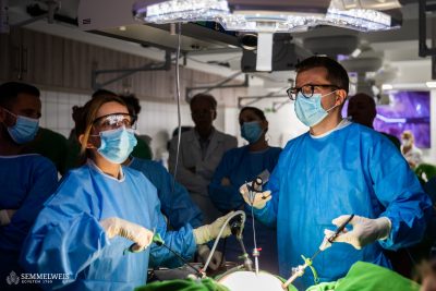
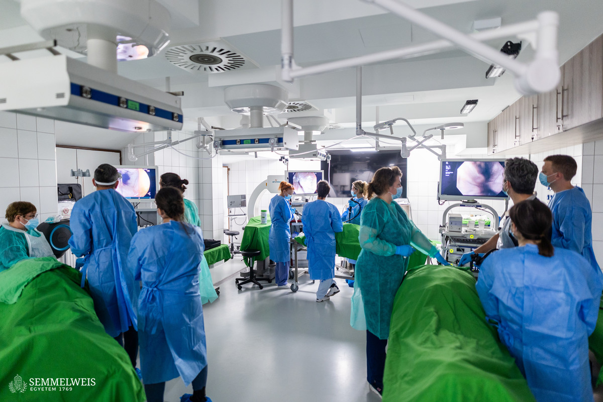
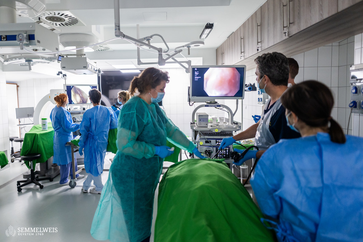
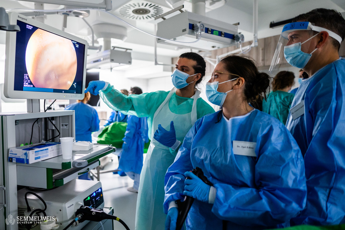
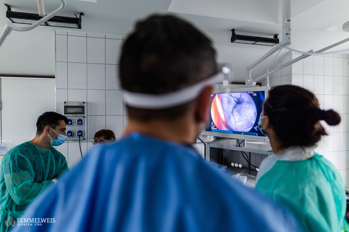
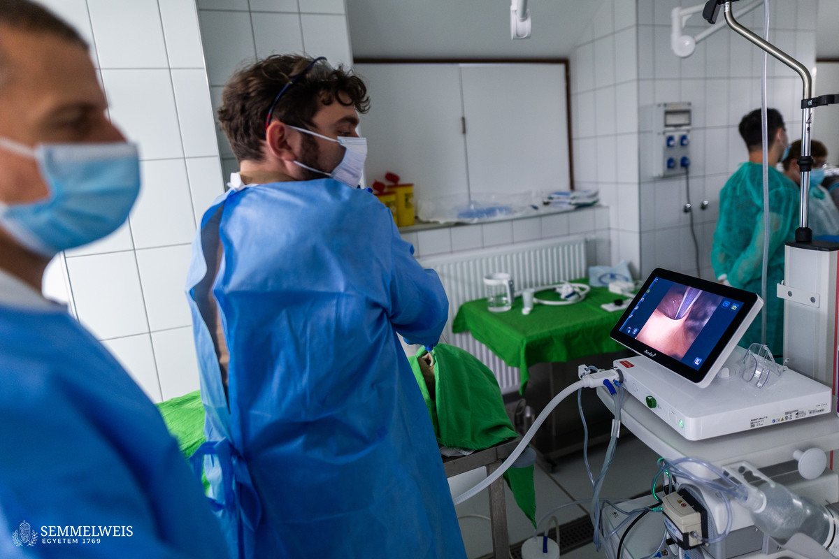
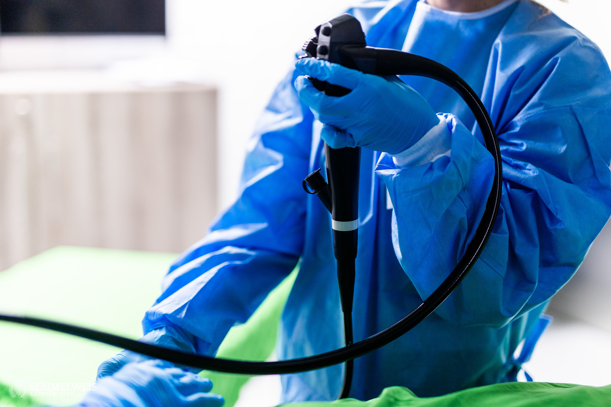
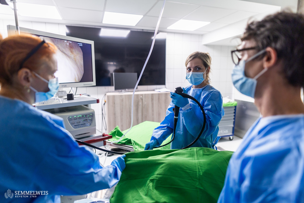
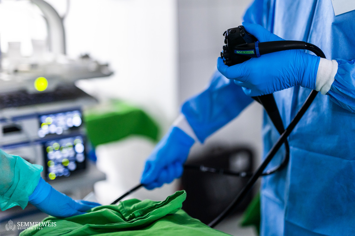
Four cadavers were available with endoscopy towers made by four different manufacturers for the morning gastroscopic intervention training. In case of each, one gastroenterology instructor discussed practical information, after which they presented an examination, and then the candidates took their turn. There was plenty of time to stop at the points which usually pose a challenge during real-life examinations, emphasized Dr. László Barkai, noting that such a case is gastroscopy, when the pharynx is passed through, during which procedure a delay is difficult for patients to tolerate. The participants received feedback immediately after the exercise. When both candidates finished at the given cadaver station, they got rotated so that by the end of the morning everyone could experience practicing on endoscopes that differ somewhat in their grip, displays and other characteristics, added Dr. László Barkai.
Gallery
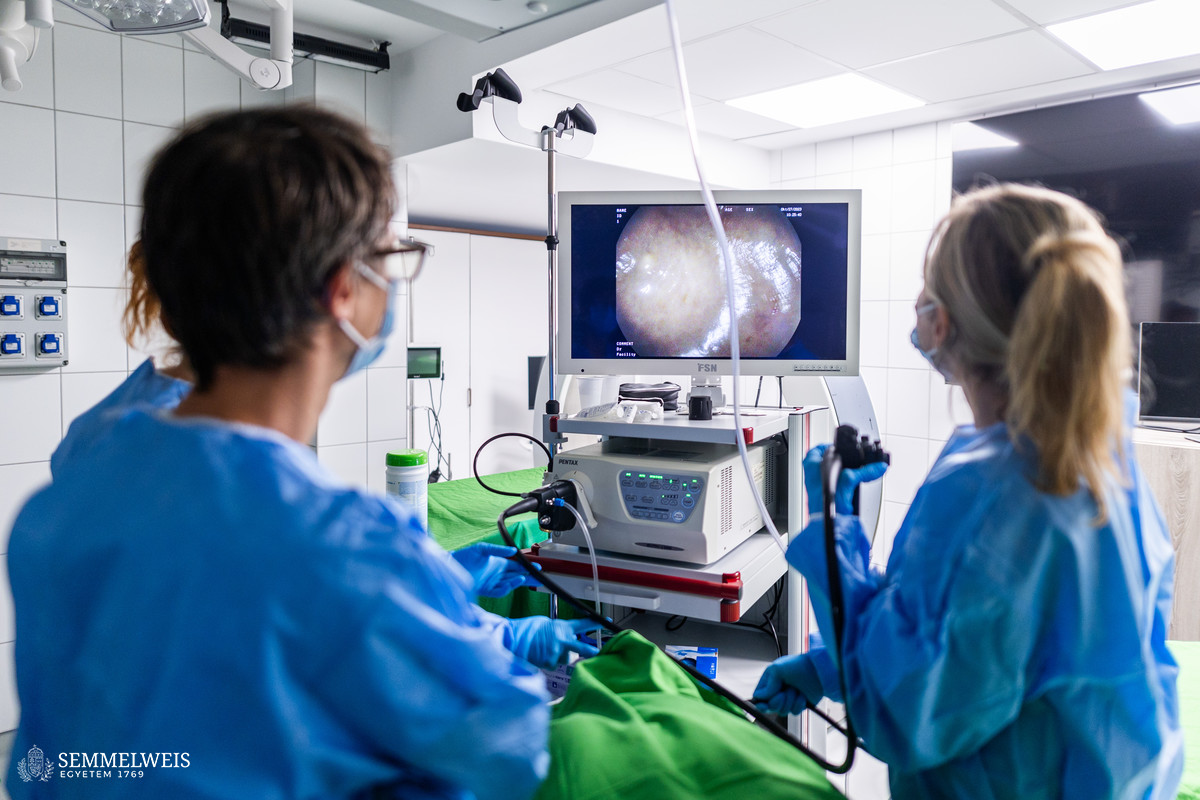
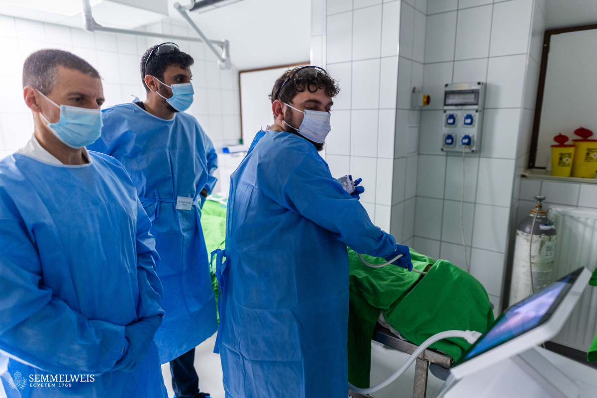
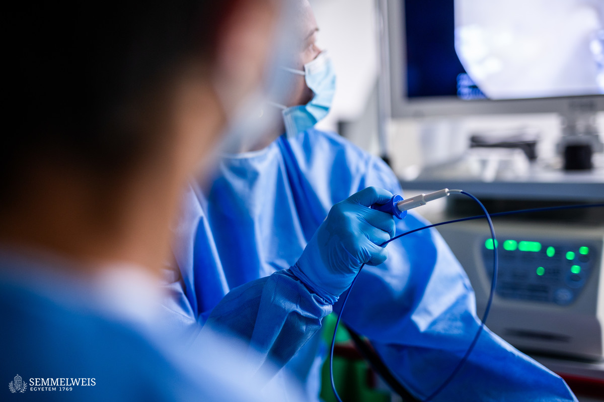
In the afternoon, the candidates could practice colonoscopy at three stations with three different types of endoscopy towers attached to three cadavers. This program also started with a short theoretical introduction, the gastroenterologists explained the characteristics, goals, and conditions of a quality colonoscopy, and they presented the examination, which was followed by the participants’ turn in a rotating system, explained Dr. László Barkai. This is a lengthy examination, so during the course it was particularly beneficial that you could spend time in calm conditions, think about the anatomy, rotate the endoscope, and practice the movements at every difficult bend or loop, he emphasized. Of course, everyone received feedback after this procedure too. According to him, the opportunity for performing an endoscopy procedure on lifelike anatomical models for a sufficient amount of time while receiving immediate feedback from the teaching gastroenterologists was particularly beneficial during the course.
The cadaver courses connecting multiple specializations and realistically depicting different clinical situations take place in the Interdisciplinary Cadaver Operating Room of the Department of Anatomy, Histology and Embryology, which was handed over at the end of 2022. In the spring of 2023, in addition to the advanced airway management training, which has been organized with great success for several years and always with the maximum number of participants possible, a gynecology-focused course was also held, which aimed to teach how to avoid serious complications that may occur during hysterectomy. The autumn-winter season was opened by a gastroenterology endoscopic course organized by the joint initiative of several university departments, which was followed by a liver and pancreatic surgery training. In addition to the advanced airway management course, the program also included a training in dental implantology, another one enabling the practice of explorations used during surgical interventions on the upper extremity, as well as a beating heart coronary artery surgery course. In the spring of 2024, in addition to the re-running of the previously organized courses, pulmonary endoscopy, ultrasound-guided follicular puncture among the assisted reproduction technologies, and treatment of upper extremity nerve injuries were added to the palette of the accredited cadaver training program.
Anita Szepesi
Translation: Mária Sánta
Photo: Bálint Barta – Semmelweis University