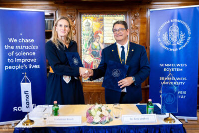
Simpler, more objective cardiac CT scan results can be obtained with the help of a medical records platform developed in collaboration between Semmelweis University and Neumann Medical Ltd. and Arineta Ltd., a cardiac CT scanner manufacturer in Israel.
As a result of the project with the identification number 2019-2.1.10-TÉT-IL-2019-00001, closed on 31 December 2021, a software was developed that can provide an answer to the problems of data collection and decision support by standardising the cardiac CT medical record workflow.
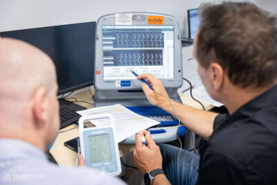
Coronary CT has become an important routine diagnostic tool in cardiology in less than 20 years, thanks to the rapid advances in technology. The period of continuous innovation is not yet over, and many more opportunities are just beginning to be exploited. However, the fast technical development in medical recording require a large amount of clean, standardised and easily assessable data, which is currently not available on the market. The practical difficulty of data collection is caused by the fragmentation of the workflow, among many other technological and medical problems. At present, at least two software tools are used by the interpreting physician to evaluate and document a cardiac CT scan properly. The images are evaluated with the “workstation” software, while the documentation is done on a completely separate interface, the hospital software. Evaluation and documentation are thus linked by an additional human work phase, which limits the amount of data recorded and results in subjective selection, in addition to the fact that the recorded image elements and clinical data can only be linked with a considerable amount of extra work.
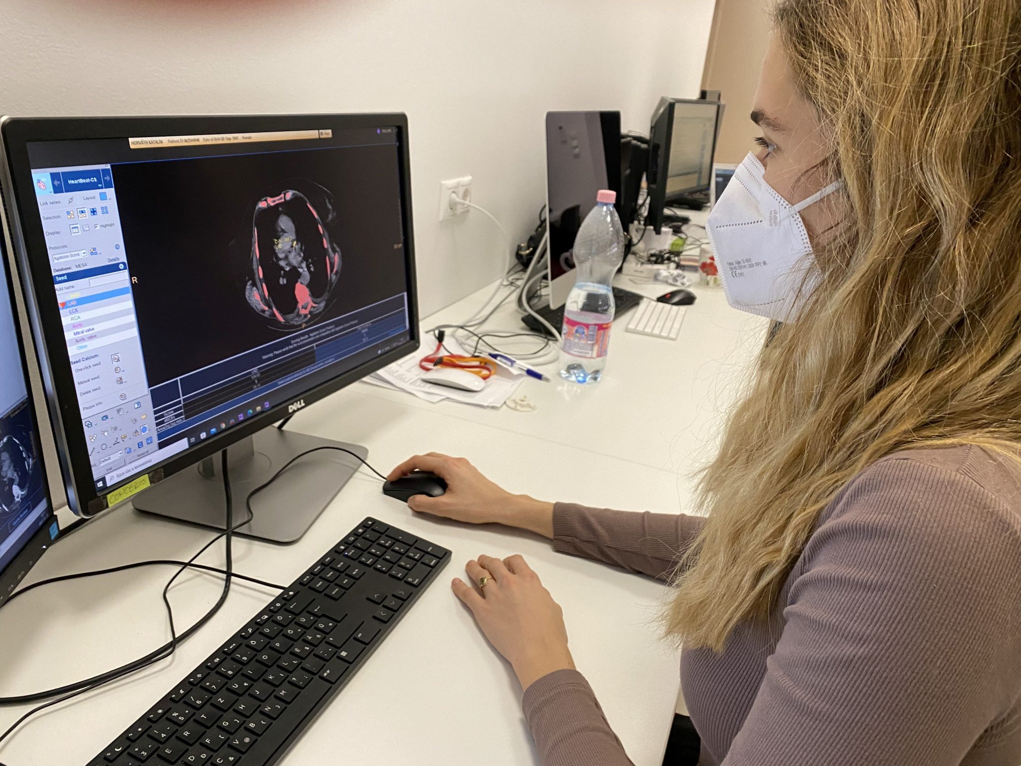 The medical records platform created as a result of the project is about connecting workstation functions (image analysis and evaluation) with clinical care needs. This will be supported by structured data entry. During the work process, the CT image is opened with the radiology image analysis workstation software that is part of the system. Here, the image is evaluated and an artificial intelligence training database is generated from the captured data in the background, without any further interaction.
In the course of the process, the relevant information recorded is automatically transferred to a structured patient care form, automatically filling in the sections of that form. The developed medical records platform allows that with minimal documentation work, the process of medical recording equals to the image evaluation itself, as the structured form also ensures the automatic generation of a medical records report.
Thus, the structured data collection is integrated into the workflow, without generating additional tasks for the doctor, and is recorded in such a quality that it is suitable for machine learning and statistical analysis without further cleaning and filtering. The accumulated data is used to train an artificial intelligence algorithm in the system that can perform part or all of the image analysis. The decision support system thus created will both answer the professional challenges outlined above (clean data, unified workflow, artificial intelligence-based decision support) and significantly simplify the practitioner’s work, including the process of medical recording and image analysis.
The project was funded by the National Research, Development and Innovation Fund of the Ministry of Innovation and Technology with a total grant of HUF 195 661 864, under the 2019-2.1.10-TÉT-IL program.
Photo (illustration): Medical Imaging Clinic