 The study involved 41 specialists from three leading spine surgery centers in Hungary. Using questionnaires, the researchers assessed the perceived advantages of life-sized 3D models created from medical imaging data compared with conventional imaging techniques such as X-rays or CT and MRI scans.
The greatest benefits were seen in surgical planning and patient communication, especially in cases where abnormal spinal anatomy or previous interventions — such as implanted metal hardware — made anatomical structures difficult to study.
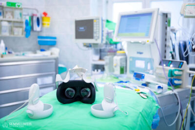
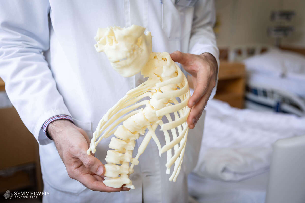
A key advantage of the 3D models is that, instead of relying solely on images displayed on a screen, surgeons can examine a physical, tangible replica of the spine. This allows them to inspect the anatomy from every angle and may facilitate a better understanding of unique anatomical variations and complex pathologies.
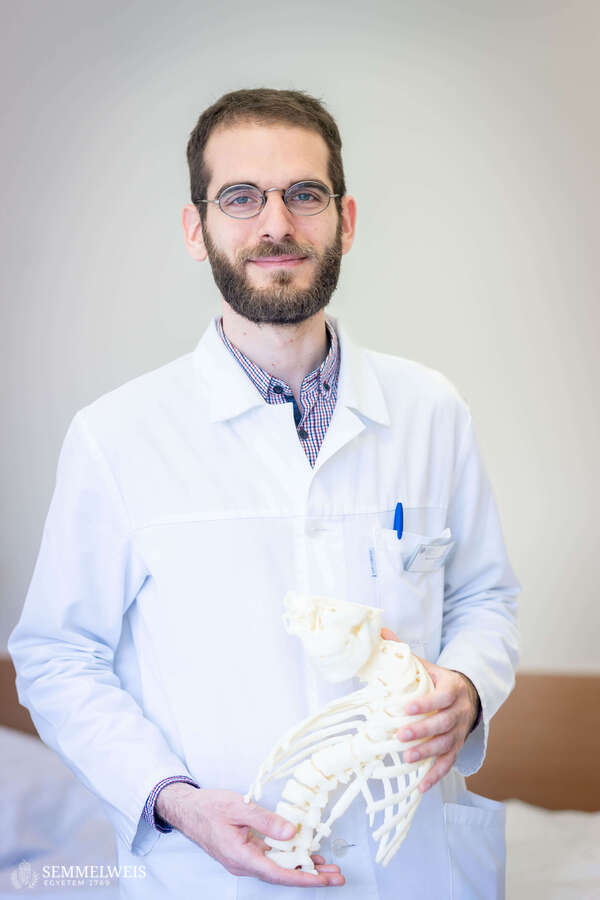
“In highly unique anatomical situations, every case represents a new learning process for the surgeon. Previous general anatomical knowledge alone is often not sufficient. In such situations, it is particularly valuable not only to see a two-dimensional image of the spine but also to hold it, rotate it, and examine it physically before surgery. The models can even be used for rehearsal procedures, allowing surgeons to explore different technical aspects of the operation, including drilling into the model,” said Dr. Péter Éltes, MD, PhD, supervisor at the School of PhD Studies at Semmelweis University, senior author of the study, and spine surgeon at the National Center for Spinal Disorders.

Before 3D-printed spine models can be routinely used in surgical practice, healthcare institutions must meet strict quality assurance and regulatory requirements. For this reason, such models are currently produced mainly for research and educational purposes. Experts believe, however, that the technology could also play an important role in patient communication in the future. A tangible model may help patients and their family members better understand the nature of the condition and the planned surgical procedure.
 “We could say that patients may literally hold a replica of their own or their child’s spine, a model of a complex congenital spinal deformity. Physicians can then explain the planned intervention in detail using the model. This represents a major step forward compared to CT or MRI scans, where we can only show two-dimensional segments. Moreover, understanding such images is often difficult for people without medical training,” emphasized Benjámin Hajnal, PhD student at the School of PhD Studies at Semmelweis University and first author of the study.
Another interesting finding was that surgeons’ opinions of the 3D models were largely independent of their experience level or specialty. Spine surgeons with decades of clinical experience found the models just as useful as their younger colleagues. In addition, 80 percent of the participants had never previously used a 3D-printed anatomical model in clinical practice. These findings suggest that the technology may have broader applications in the preparation of complex spine surgeries.
Ádám Farkas
Photo: Boglárka Zellei – Semmelweis University