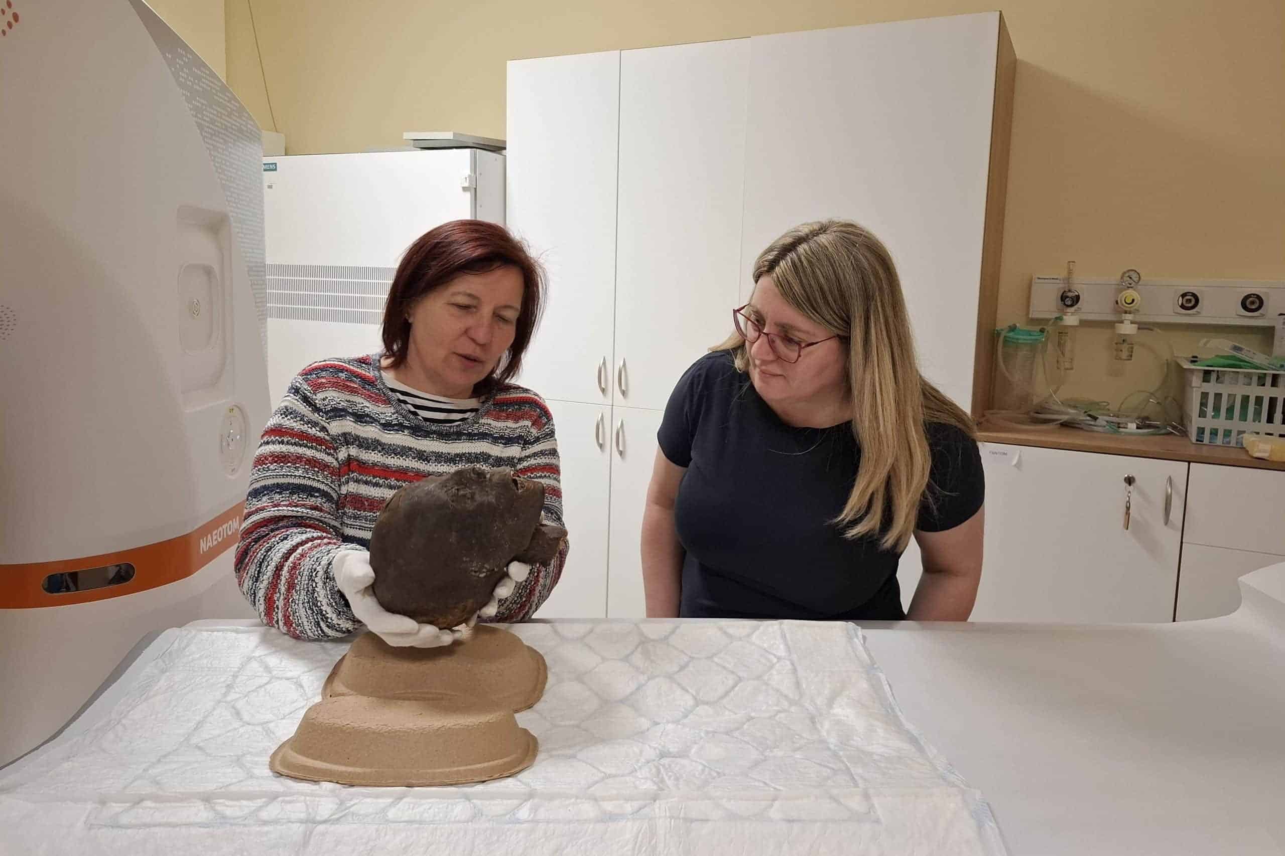
In accordance with standard clinical practice, the examination of the mummy remains was conducted outside of patient examination hours, at night. This technology is particularly effective at analyzing complex, multilayered materials, including the non-destructive, detailed analyses of mummified human remains. “The aim of the examinations is to obtain as accurate a picture as possible of the internal structure of the remains, any abnormalities, and the preservation techniques used,” said Dr. Ibolyka Dudás, Chief Clinical Physician at the Department of Radiology and Head of the working group for post-mortem imaging.
Over two-thousand-year-old finds under CT scan
The Egyptian mummy remains currently under examination were added to the collection of the MNMKK Semmelweis Museum of Medical History around the time of its founding. In recent years, the finds in question have been repeatedly subjected to various imaging and multidisciplinary examinations, including conventional CT scans. However, due to technological constraints, these did not allow for a sufficiently detailed assessment. Six specimens were subjected to radiocarbon (C14) dating, but only three yielded measurable results. Based on this, the oldest remains can be dated to between 401 and 259 BCE, meaning they are more than 2,300 years old. As part of the current series of examinations, this specimen and all of the museum’s other Egyptian mummy remains have undergone new analysis.
Novel opportunities for more accurate diagnosis
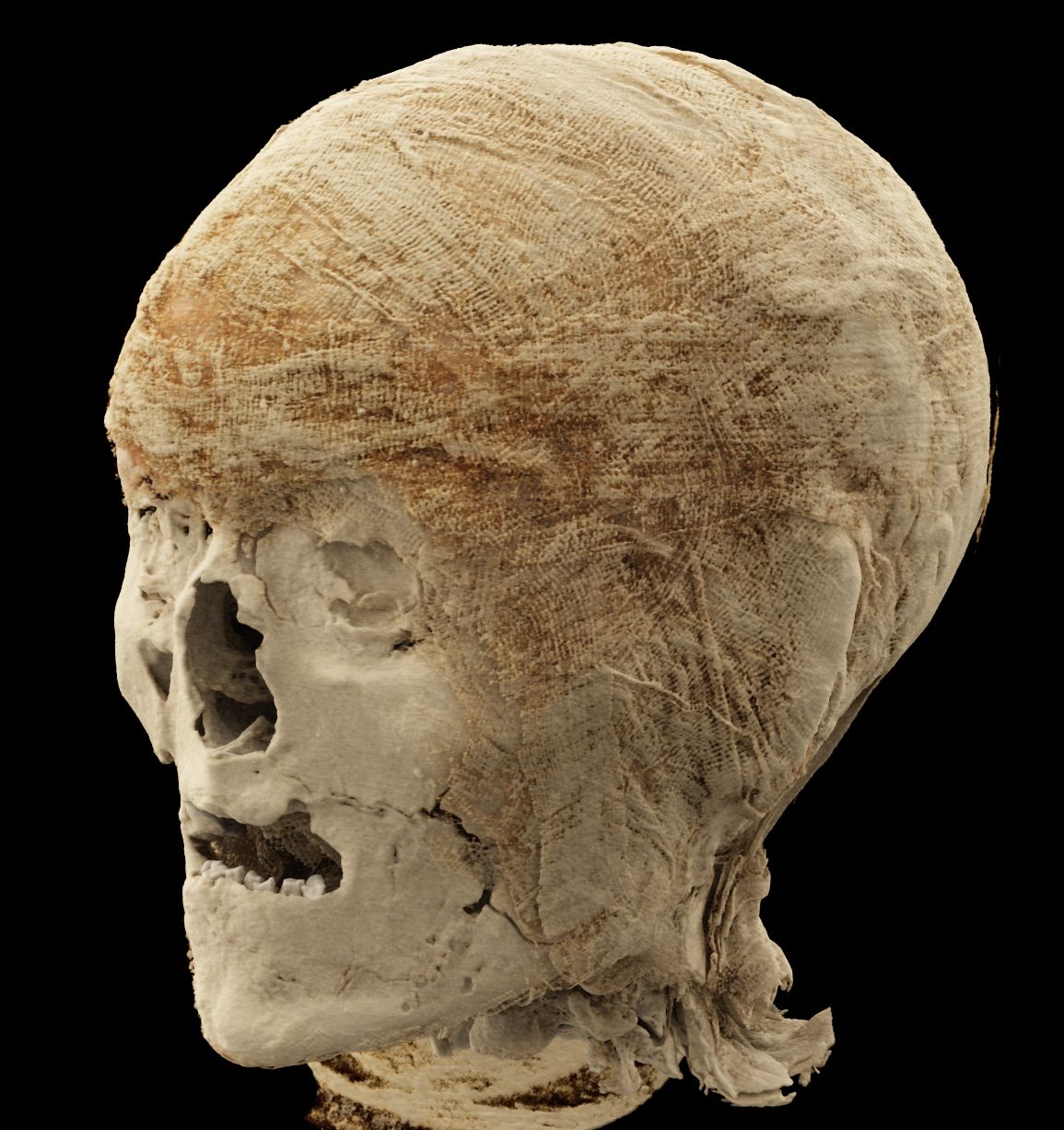
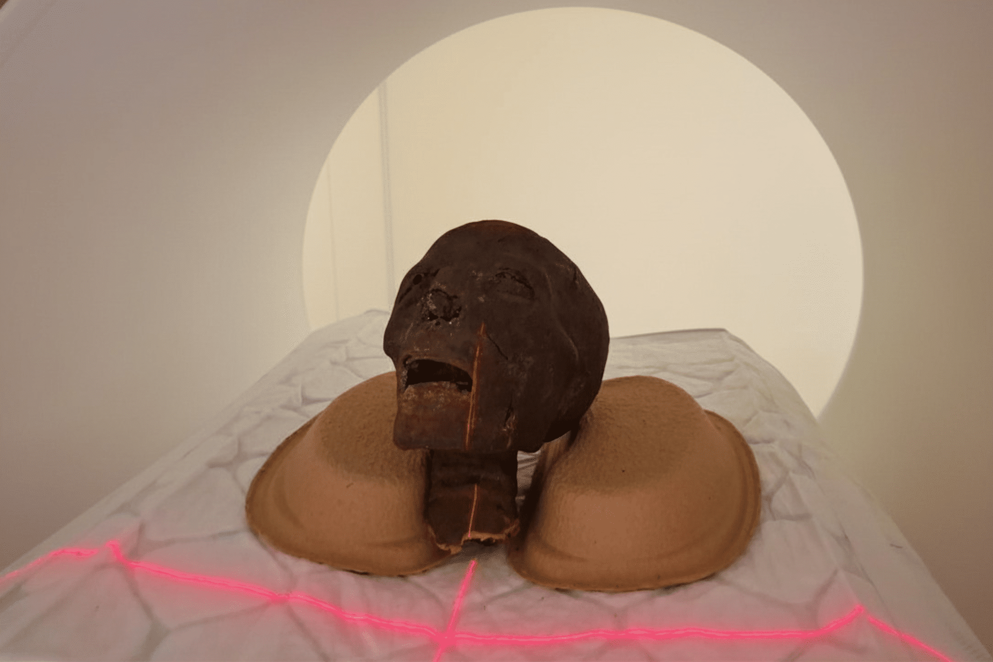
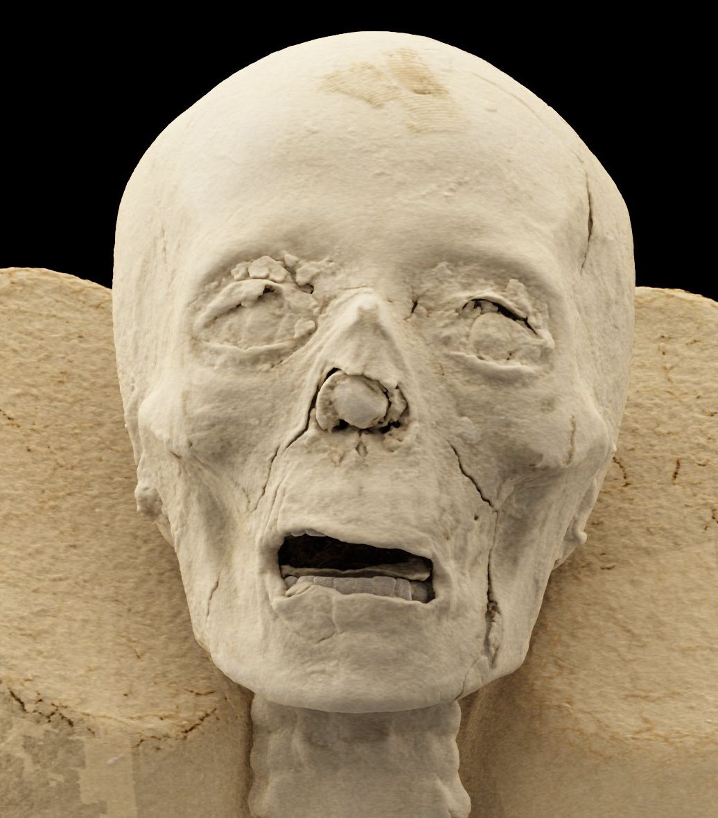
The recent high-resolution CT scans allow for a more detailed examination of the teeth and skull sutures of the two mummified heads. This could lead to a more accurate age determination and lay the groundwork for future high-precision, detailed 3D reconstructions, as well as potential facial reconstructions of the skulls.
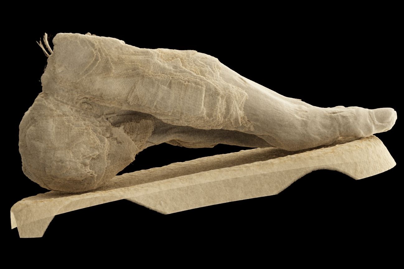
In the case of a previously examined left lower limb, it was not possible until now to establish a definitive diagnosis; however, based on the new images, several possible interpretations have emerged. The current examinations indicate that the individual may have suffered from osteoporosis; however, the exact cause – whether due to age-related factors or a pathological process – requires further, detailed analysis.
An examination of the second lower left limb revealed that the remains likely belong to a young individual. The exact age is still being determined, but this is the first time such detailed imaging data has become available for this find.
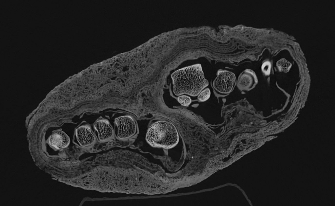
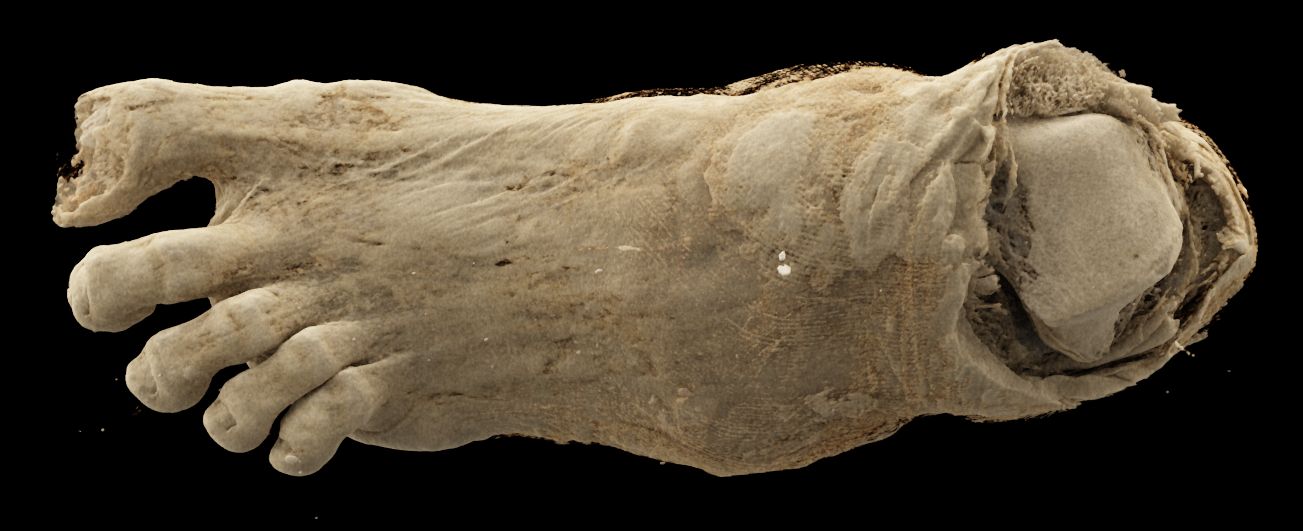
The examination of a set of remains – previously interpreted merely as a mummy bundle – yielded particularly noteworthy results. When the find arrived at the museum, in the absence of imaging tests, it was initially identified as a human head and later, possibly, as a bird mummy. However, a previous CT scan clearly showed that the find is, in fact, an adult foot. The current imaging analysis aims to determine the extent to which the textile remnants can provide insight into the mummification technique, the age of the mummified individual, and any illnesses the individual may have had. The current images clearly reveal the different layers of the bandage and show their different structural characteristics. These findings may lay the groundwork for further historical and technological investigations. The remains were presumably part of a complete mummy, but the reason and time of the dissection are currently unknown.
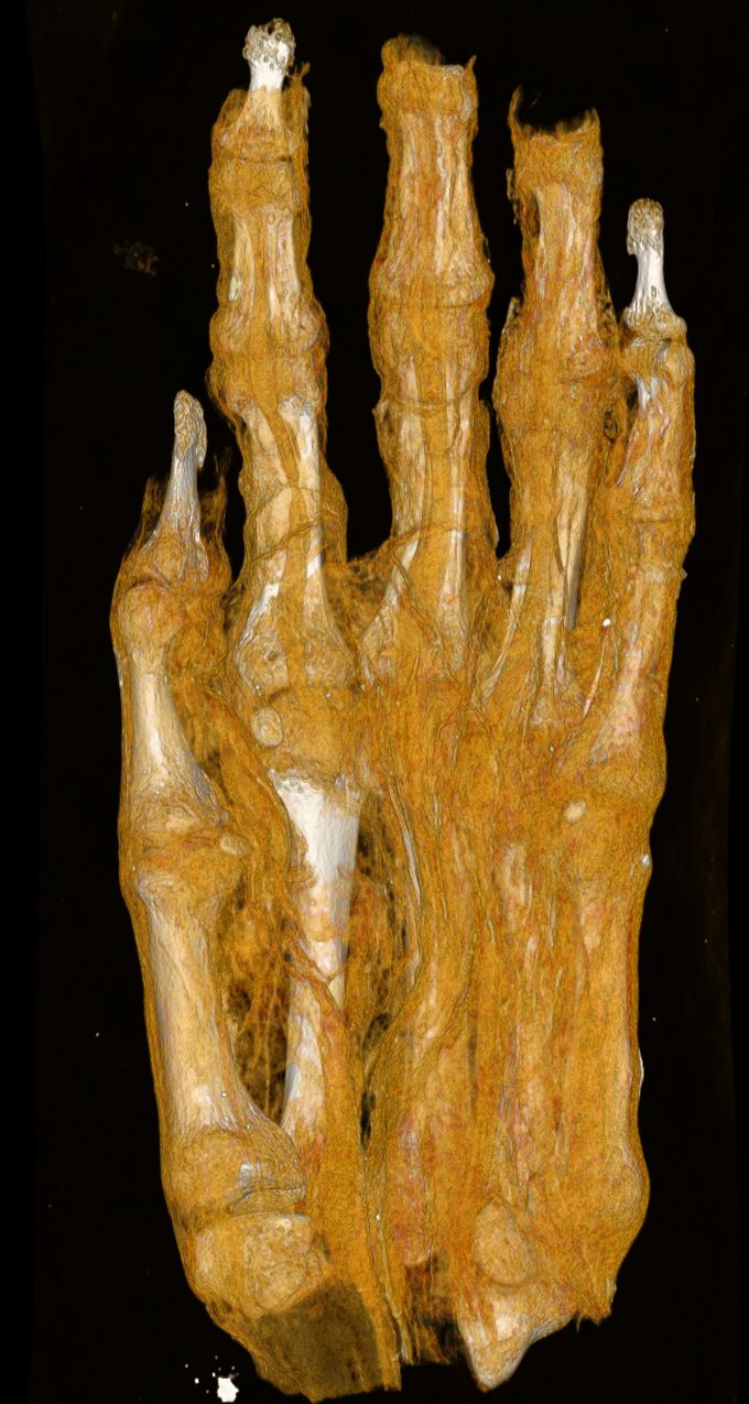
The analysis of the mummified hand included in the study can also offer valuable information. Based on the size, development, and morphological characteristics of the bones, researchers aim to determine whether the remains belonged to a child or an adult. They may also be able to estimate the individual’s sex and age.
Cutting-edge technology in the service of mummy research
“The remains had previously been examined by a research team, but the current images provide a more detailed view than ever before and are expected to yield new, scientifically valid findings regarding the remains that have been preserved in the collection for decades,” emphasized the collection’s curator, Chief Museologist Krisztina Scheffer.
A detailed evaluation of the images is currently underway. Researchers expect the data analysis to provide new insights into the mummies’ lives and state of health, as well as the mummification process.
Gallery
“Based on the results so far, it is evident that modern imaging technology opens up new perspectives in mummy research. It can reveal information hidden in finds that are thousands of years old without damaging them,” added the chief curator.
Enikő Szvák
Translation: Judit Dőtsch
Image credit: Medical Imaging Center (OKK), MNMKK Semmelweis Museum of Medical History