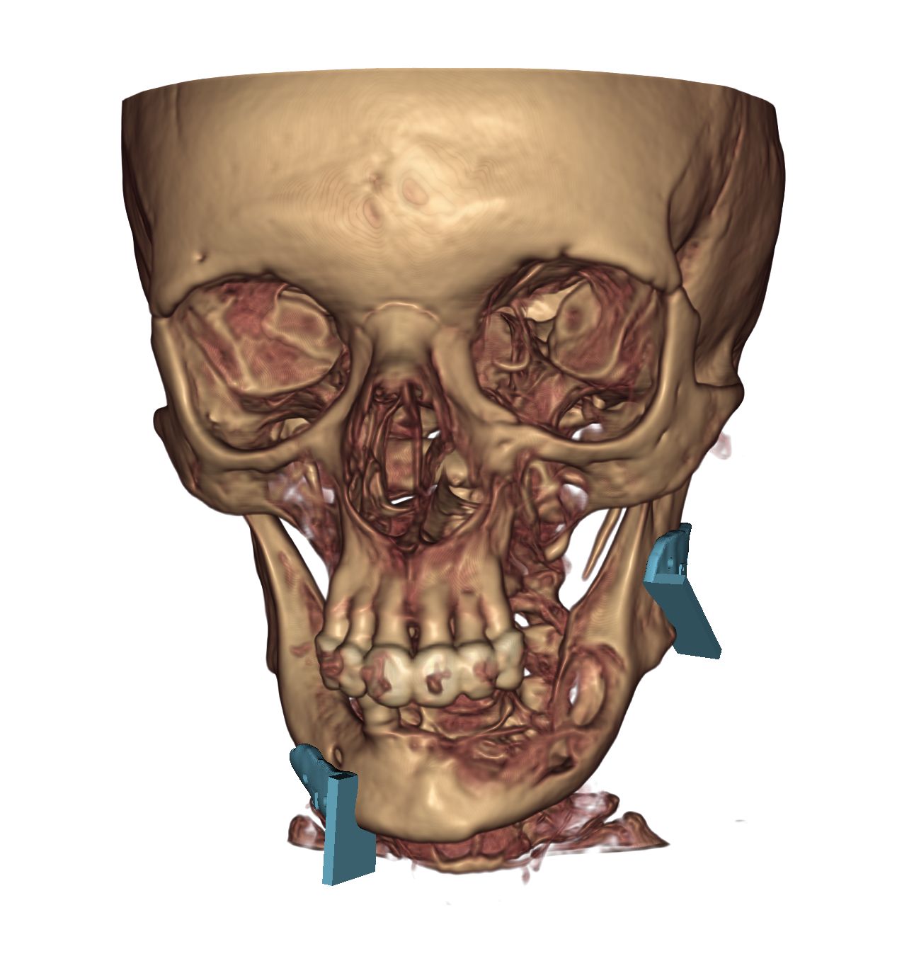
Most commonly used after the removal of malignant tumors of the oral cavity and certain benign tumors, the jaw reconstructive surgery at Semmelweis University can even restore the face and the chewing function to its original state. The surgeries are performed in close collaboration between specialists from two university departments (plastic surgeons and oro-maxillofacial surgeons), who also jointly plan the procedure.
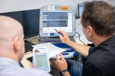
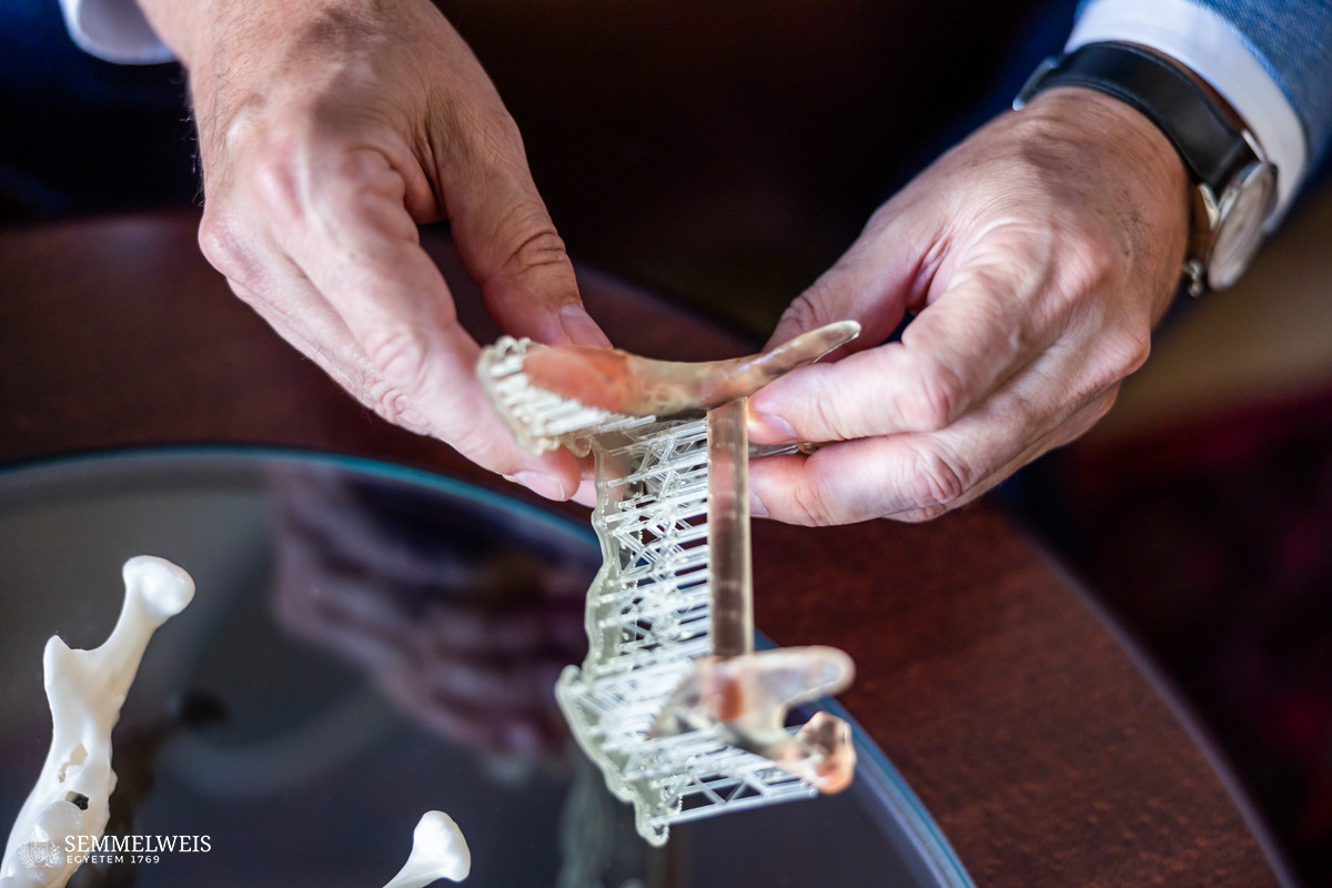
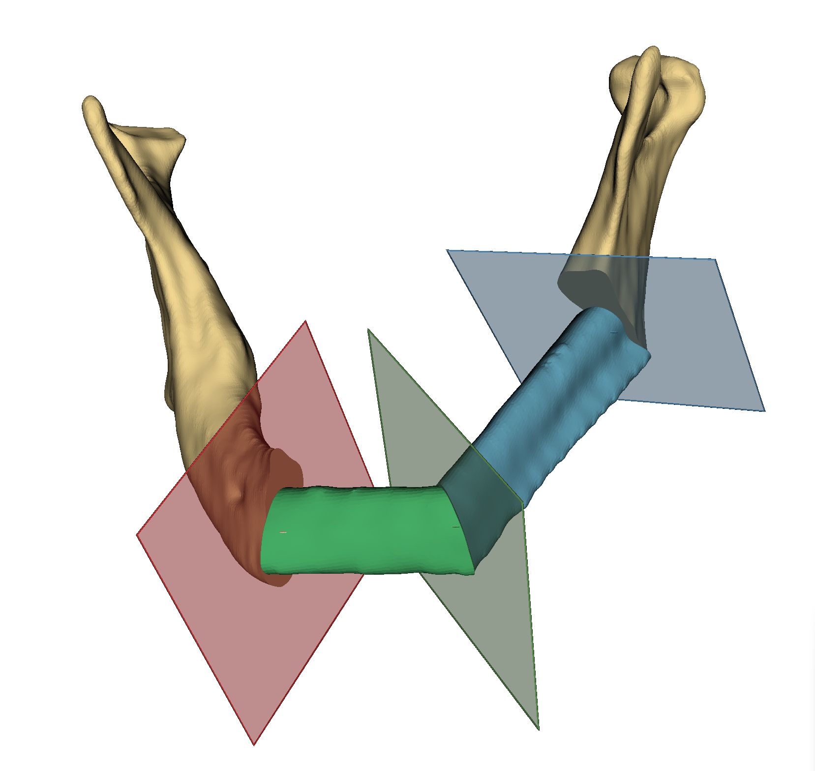
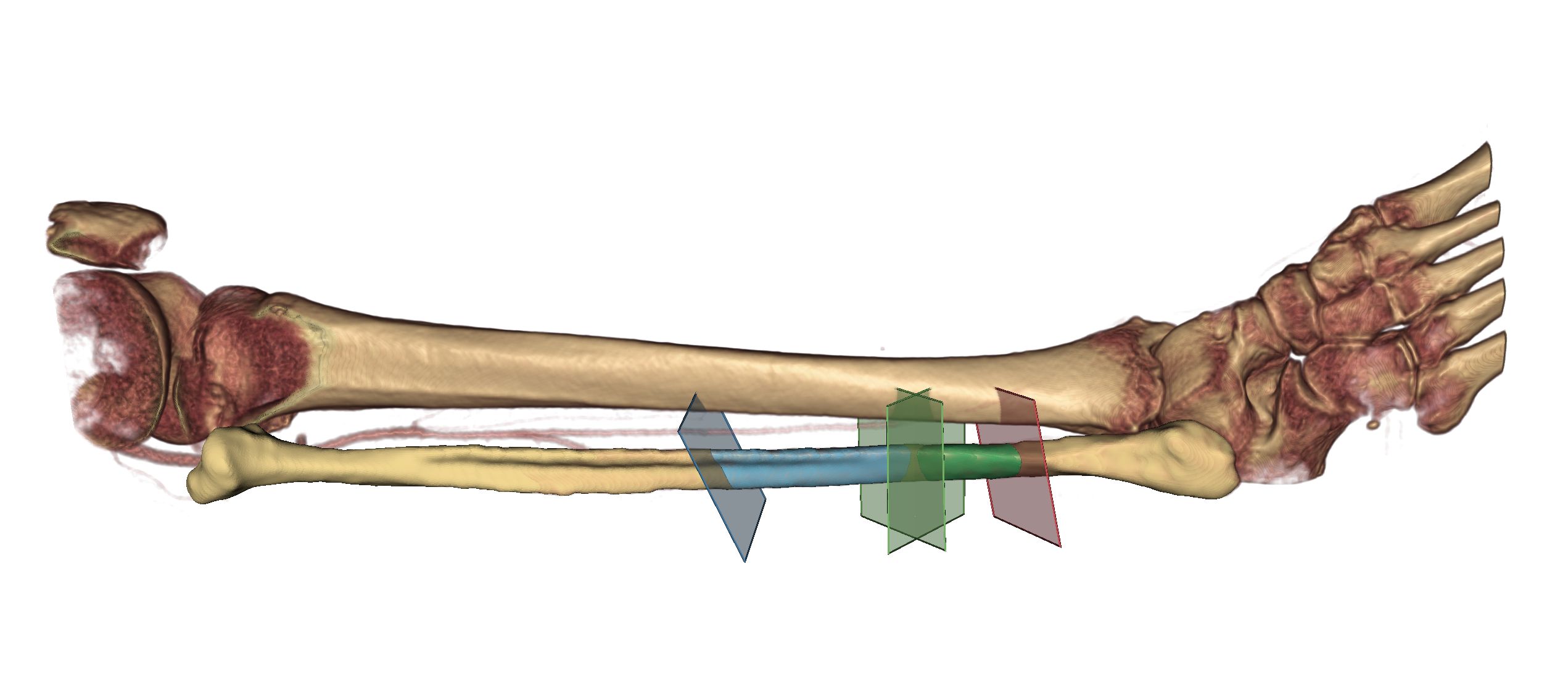
 “With imaging, software-based design, and 3D technology, we can simulate in advance what we are going to do in the operating room. By creating virtual plans, we can model all the details of the operation in advance, from how much of the shin bone to remove, how many pieces to cut it into, and where to break the bone,” said Dr. Tamás Würsching, a specialist at Semmelweis University’s Department of Oro-Maxillofacial Surgery and Stomatology, who is in charge of the 3D planning process. He pointed out that before this procedure became available, measurements had to be made at the operating table, which made the surgery significantly longer, while the operating time is the most precious aspect.
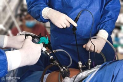
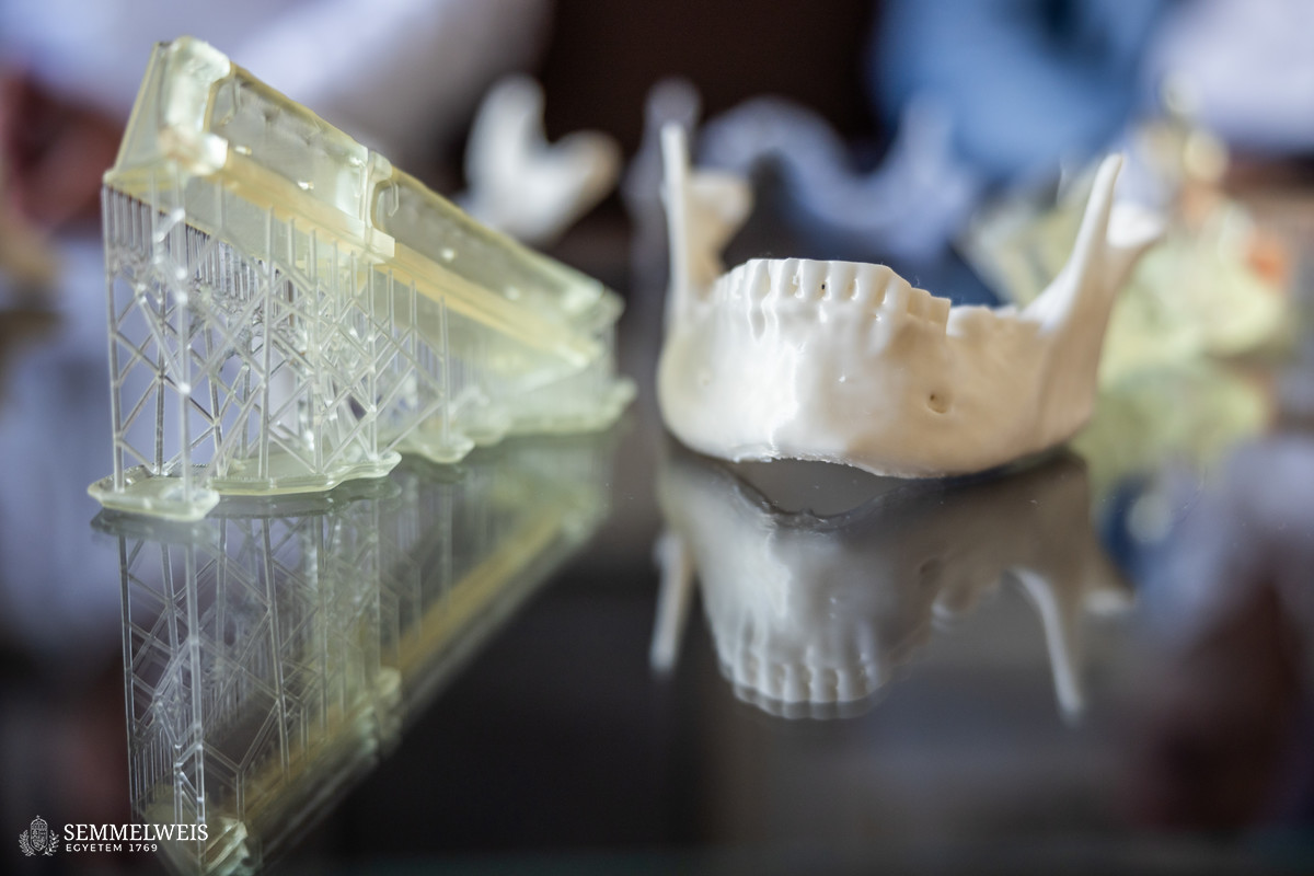
The most suitable bone to replace the jaw is the fibula, a long bone on the outside of the lower leg. Because of its stability, shape, and hardness, dental implants can be placed in it, so that the original chewing function can be preserved after the tumor is removed. In the event of uncomplicated healing, this method does not affect walking either, allowing a full life to be lived.
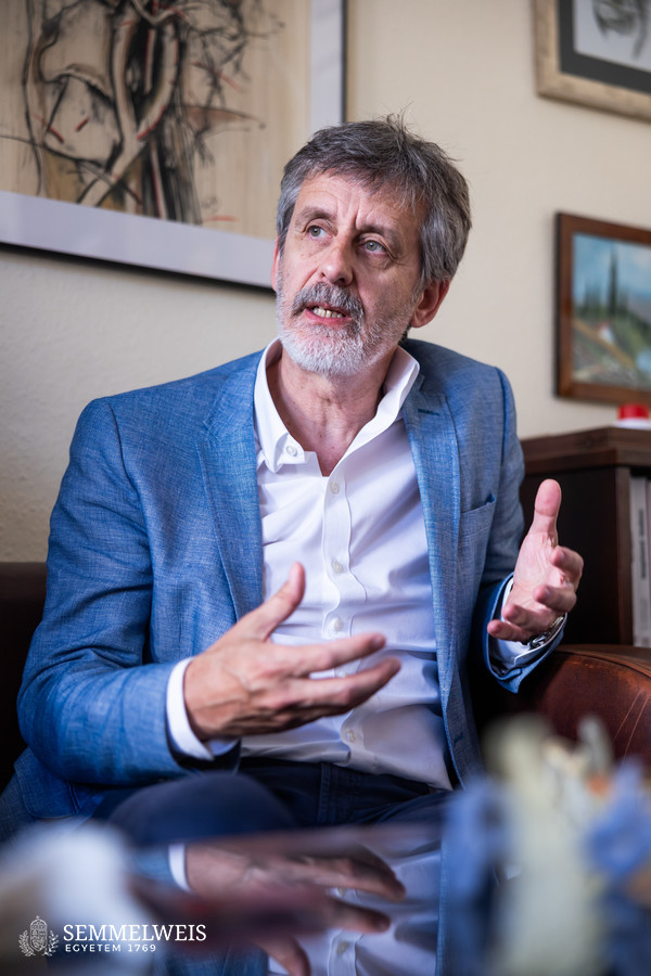 “If the soft tissues surrounding the patient’s jaw are intact after the tumor is removed, only the bone has to be replaced. If the surrounding tissues (e.g. skin) also need to be replaced, then the piece of the fibula is removed and transplanted together with the soft tissues,” added Dr. Sándor Bogdán, Associate Professor and Head of Semmelweis University’s Department of Oro-Maxillofacial Surgery and Stomatology. Part of 3D design is also to determine exactly what type and size of tissue is needed for a given patient, he stressed.
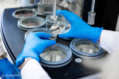
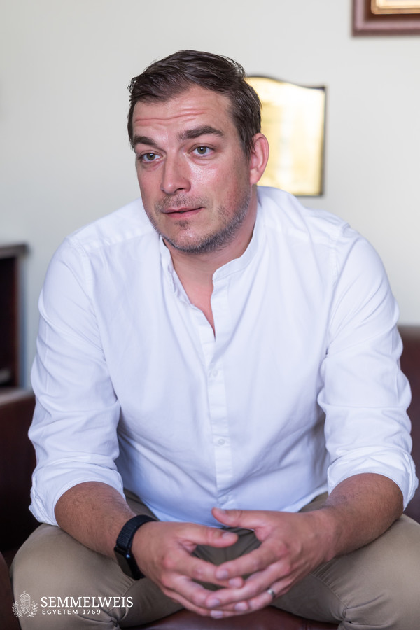 “Using the designing software, a 3D model of the lower leg is created, on which the bone section to be grafted is marked. We then use a 3D printer to create cutting templates that are applied to the fibula. In this way, we can cut through the selected parts at specific angles, which the surgeons can immediately insert in the right place, and then we also ensure the blood supply to the transplanted flap by suturing the blood vessels,” said Dr. Zoltán Klárik, plastic surgeon at Semmelweis University’s Department of Surgery, Transplantation and Gastroenterology. He added that the success of the operation depended largely on the length of time the bones to be grafted were deprived of oxygen and blood supply. After an hour, irreversible tissue damage occurs, so time is of the essence during surgery. For this reason, surgery with cutting templates that have been precisely planned in advance greatly improves the success of the operation.
This procedure is used in several centers in Hungary, and Semmelweis University has been performing jaw reconstructive surgery with this technology for three years. Every year, around fifteen patients with an oral cavity tumor who also need a mandibular replacement – as recommended by the consulting oncology specialists – are operated on.
While this form of tumor used to be common in men aged between fifty and sixty, nowadays the gender differences are disappearing, and it is not uncommon to see patients in their thirties. Risk factors for the disease include smoking, alcohol consumption, and HPV.
Gallery
Eszter Csatári-Földváry
Translation: Dr. Balázs Csizmadia
Photos, featured image by Bálint Barta – Semmelweis University; Department of Oro-Maxillofacial Surgery and Stomatology – Semmelweis University