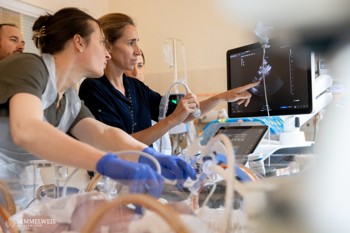
A significant proportion of babies in neonatal intensive care units (NICUs) require ventilation or ventilatory support due to poor lung function. Currently, chest X-ray is the standard for lung imaging; however, lung ultrasound (LU) provides much more reliable, detailed, and comprehensive information in many respects, emphasized Dr. Miklós Szabó, Head of the Department of Neonatology at the Pediatric Center.
 As Dr. Miklós Szabó pointed out, recognizing the significance of LU is a new trend even at an international level, which is why it is an important step that for the first time in Hungary, the center organized an international training course on the topic, led by Dr. Virginie Meau-Petit, neonatal LU pioneer and prominent international expert. “Many say that this procedure is the ‘phonendoscope of the future’, that is, LU should become as integral to the daily routine of pediatricians or neonatologists as the medical instrument is regarded as the well-known symbol of medicine,” the head of the department noted.
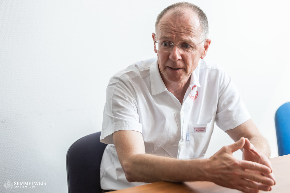
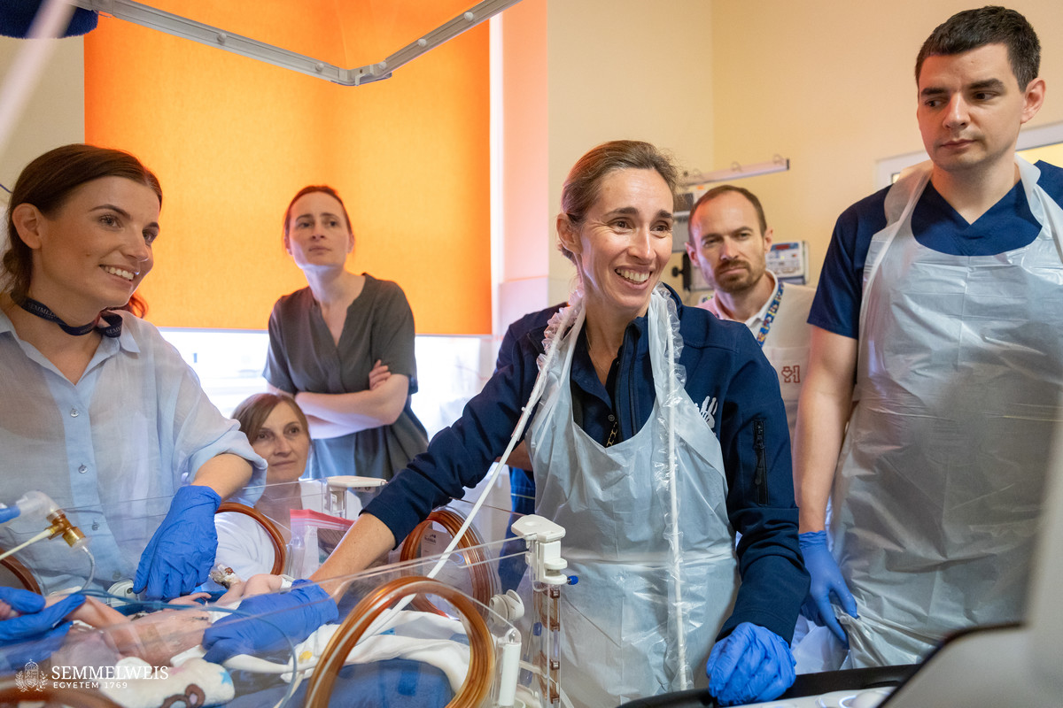
At Semmelweis University’s Pediatric Center, lung ultrasound scans have been performed in the neonatal intensive care unit for about three years, thanks to specialists who gained experience abroad. One of them is consultant neonatologist Dr. Barbara Szász, the initiator of the current training, who completed her neonatology training in London and had the opportunity to study alongside Dr. Virginie Meau-Petit, the invited lecturer of this workshop.
“When I graduated from university ten years ago, we were taught that the lungs could not be examined with ultrasound, and this was also the international consensus,” said Dr. Barbara Szász, describing how novel the approach was, and how it had started to change in the last five to ten years.
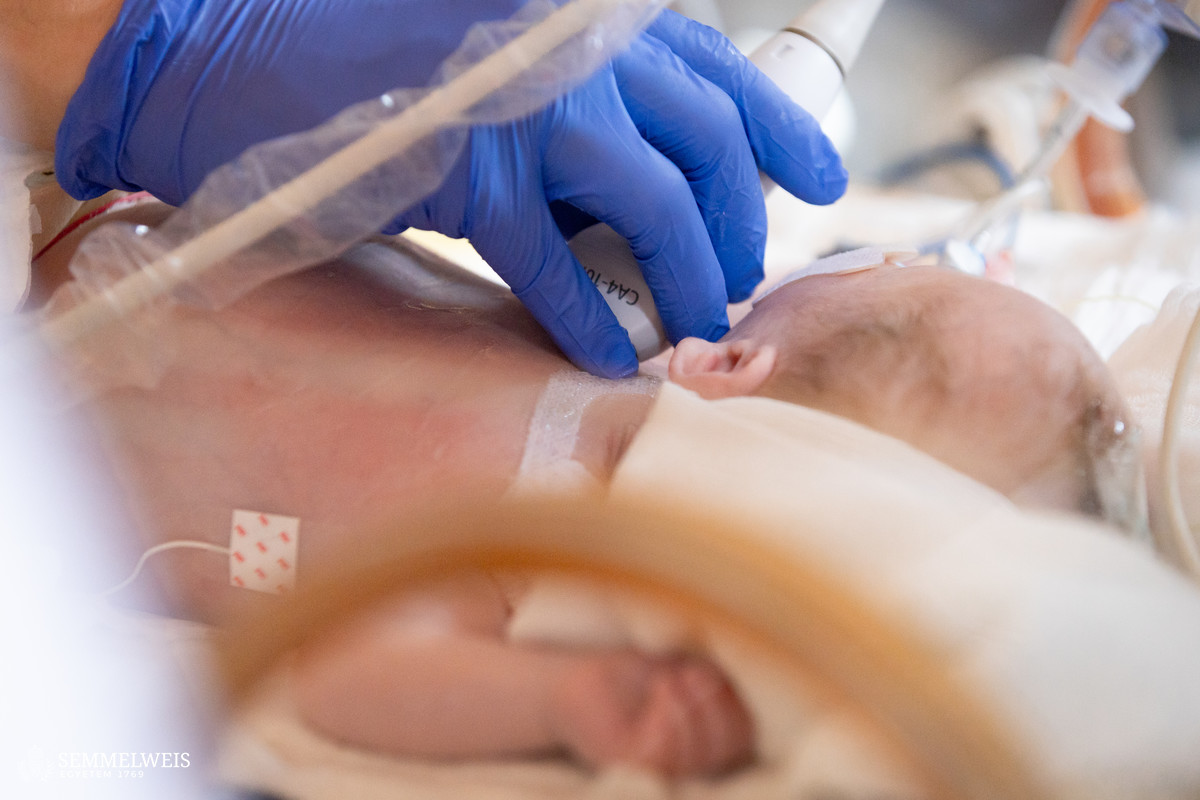
As she pointed out, the main advantages of ultrasound scanning are its easy accessibility, point-of-care (bedside) testing opportunity, swiftness, the fact that it allows for personalized medicine and offers a real-time response to diagnostic questions, without exposing the newborn to radiation. It can be particularly helpful if a sudden health deterioration requires quick decision-making, for example, it may be even life-saving when diagnosing pneumothorax.
Lung ultrasound can also facilitate the treatment of respiratory distress syndrome, a serious lung disease in premature babies due to immature lungs, as LU can detect earlier whether an intervention is needed, Dr. Barbara Szász explained.
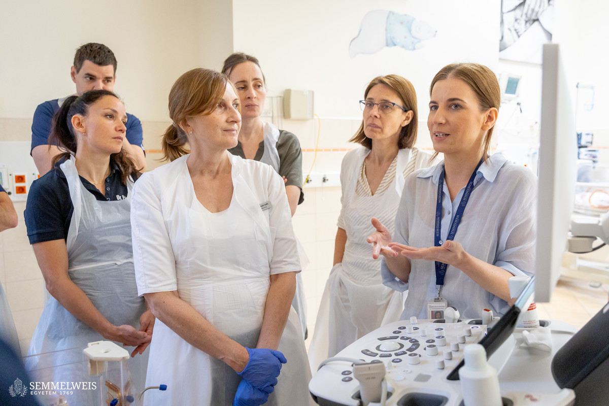
The two-day theoretical and practical training course, organized by the Department of Neonatology with the support of the Bókay Pediatric Center Foundation, was attended by the center’s staff and neonatologists from all over the country, more than thirty in total. After the theoretical introduction, participants were given bedside practical training in NICUs of the Pediatric Center and the Department of Obstetrics and Gynecology to learn the basics of lung ultrasound of neonates. Attendees will be able to consolidate their knowledge in their own departments with regular remote mentoring.
 The course was led by leading guest lecturer Dr. Virginie Meau-Petit, who had gained her extensive knowledge in level IV NICUs in Paris and London (Hôpital Necker-Enfants Malades and Evelina London Children’s Hospital). Having learned the technique in France in 2013-2014, she has been performing lung ultrasound scans on newborns for ten years and has been teaching it for six years, she told our website. “When I went to the UK afterwards, no one was doing neonatal LU at that time, only skull scans through the newborns’ fontanels. Likewise, the prevailing practice at the time was that only radiologists and cardiologists did cardiac ultrasounds, not neonatologists. As a result, our first attempt to teach neonatal lung ultrasound was a complete failure due to lack of peer-reviewed publications and recommendations,” she recalled.
Responding to the growing interest in the years after the European recommendation was published, Dr. Virgine Meau-Petit launched theoretical and practical training sessions for UK neonatologists, initially in neonatal units on site, and over time at formal trainings organized by the NeoFOCUS UK group, with last year’s course in Glasgow attracting 70 neonatologists, for example. The group’s workshops are now available not only in the UK but also in France. Participants are also offered weekly follow-up online consultations, including case discussions.
 Ultrasound is traditionally used to diagnose organs made of fluids and tissues, such as the liver, kidneys, or uterus, as ultrasound detects the boundaries between the various tissues. The uniqueness of LU technology lies in the fact that the lung is an air-rich organ, but gas is the ‘enemy’ of ultrasound. Therefore, this mode of imaging works indirectly here, i.e. it is the ratio of water to air that indicates the patient’s condition. The sicker the lung, the more fluid it contains, Dr. Virgine Meau-Petit explained.
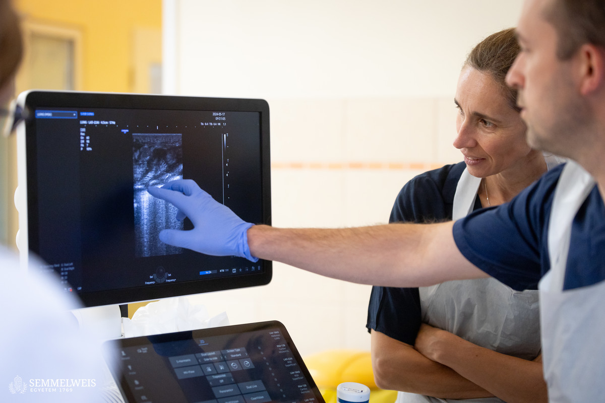
Experience shows that the advantages of this technology are even more evident in the case of newborns: the small size of the organs compared to the ultrasound transducer allows the lungs to be examined more quickly, and the entire organ can be displayed in one image, which is of better quality than with adults. “The speed and accuracy of the method represent a big leap forward in everyday care,” the expert concluded.
The technique of lung ultrasound was developed by Dr. Daniel A. Lichtenstein, an adult intensivist in France, and it took more than a decade for this originally unconventional method to gain widespread professional acceptance and to prove its validity and advantages over lung CT scans or chest X-rays. It was adapted to neonatal medicine by Dr. Daniel Lichtenstein and Dr. Nadya Yousef, whose clinical trials have provided scientific evidence of its clinical utility. The breakthrough came in 2020 with the publication by the European Society for Paediatric and Neonatal Intensive Care’s (ESPNIC) recommending the widespread use of the technology.
Pálma Dobozi, Judit Szabados-Dőtsch
Photos by Boglárka Zellei – Semmelweis University