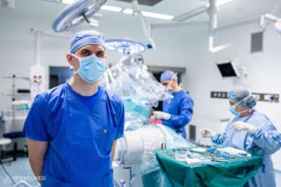
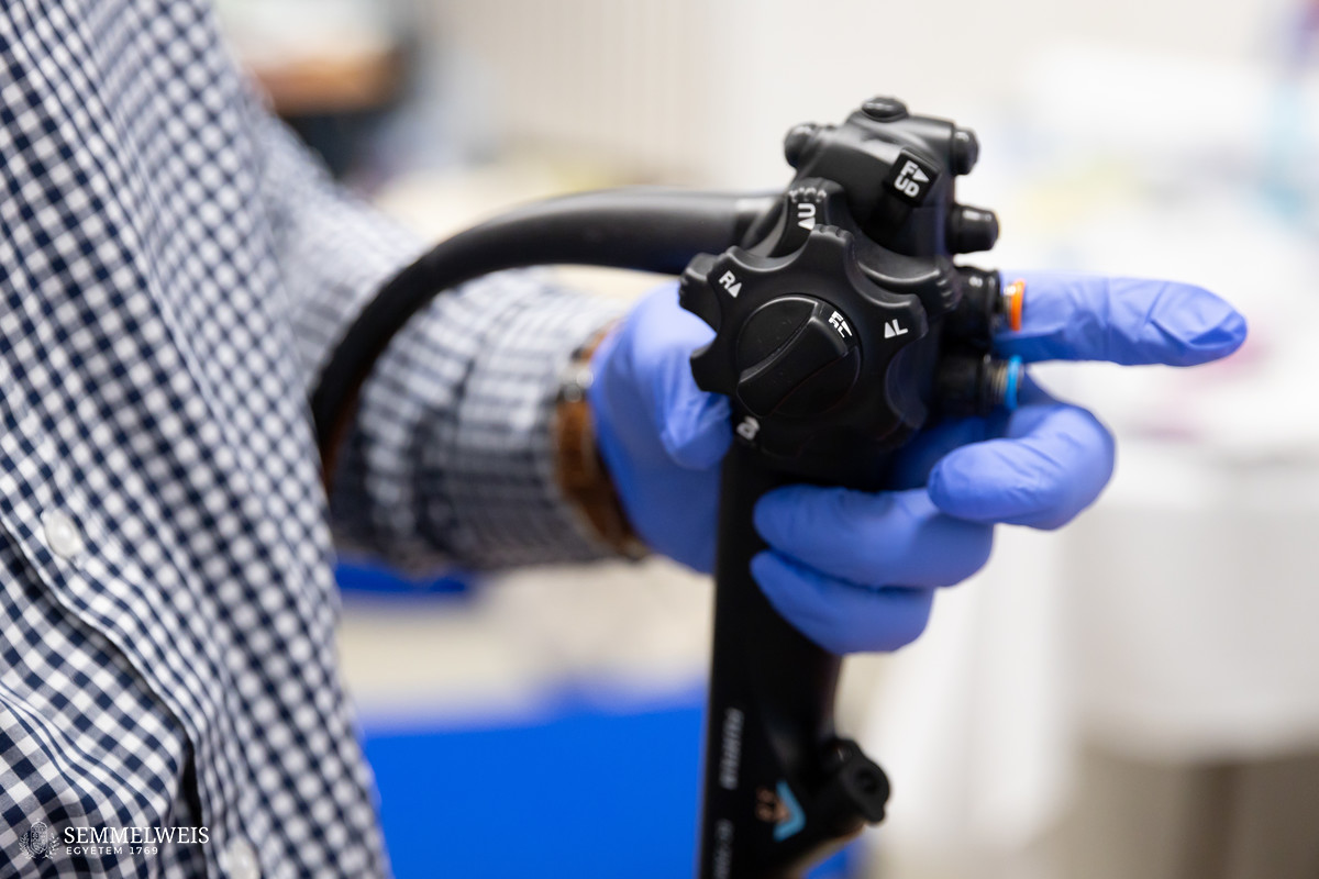
One of the cornerstones of gastroenterology training is endoscopy, which enables not only diagnostics, i.e. the detection of diseases and sampling, but also minimally invasive procedures.
According to Associate Professor Dr. István Hritz, Head of the Department of Interventional Gastroenterology, endoscopy can now be used to treat conditions that previously required surgical intervention, for example, it allows for polypectomies (i.e. the removal of polyps) and the management of gastrointestinal bleeding. Acquiring the necessary skills is an integral part of the specialized training and, consequently, of board certification as well, emphasized the chairman of the Gastroenterology Committee of Semmelweis University.
While previously the technique was mainly practiced on patients in real-life settings, which, despite expert supervision, could inevitably cause discomfort to patients, training is now assisted through simulators. These have the advantage of modeling different real-life situations while eliminating the stress related to the intervention.
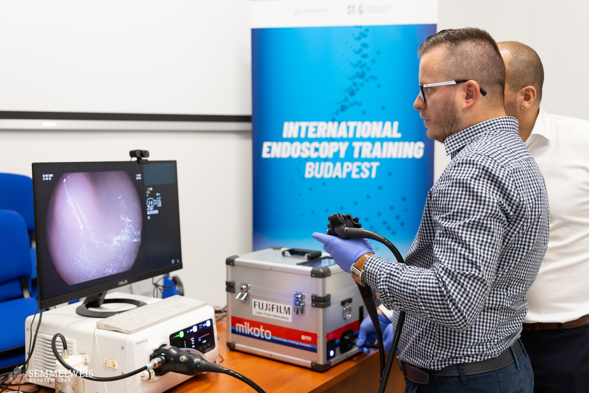
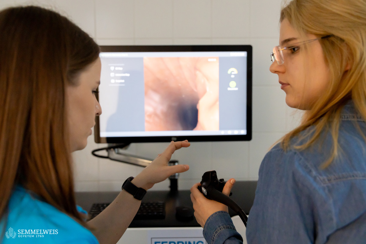
Such modeling is made possible with the Surgical Science GI Mentor computer-based endoscopic simulator, which has been available at the Department of Surgery, Transplantation and Gastroenterology since January 2024 as a courtesy of Ferring Pharmaceuticals Hungary Ltd. Its modules are suitable for mastering lower endoscopy (colonoscopy), upper endoscopy (gastroscopy), polypectomy, hemostasis, endoscopic ultrasound, as well as bile duct intervention (ERCP).
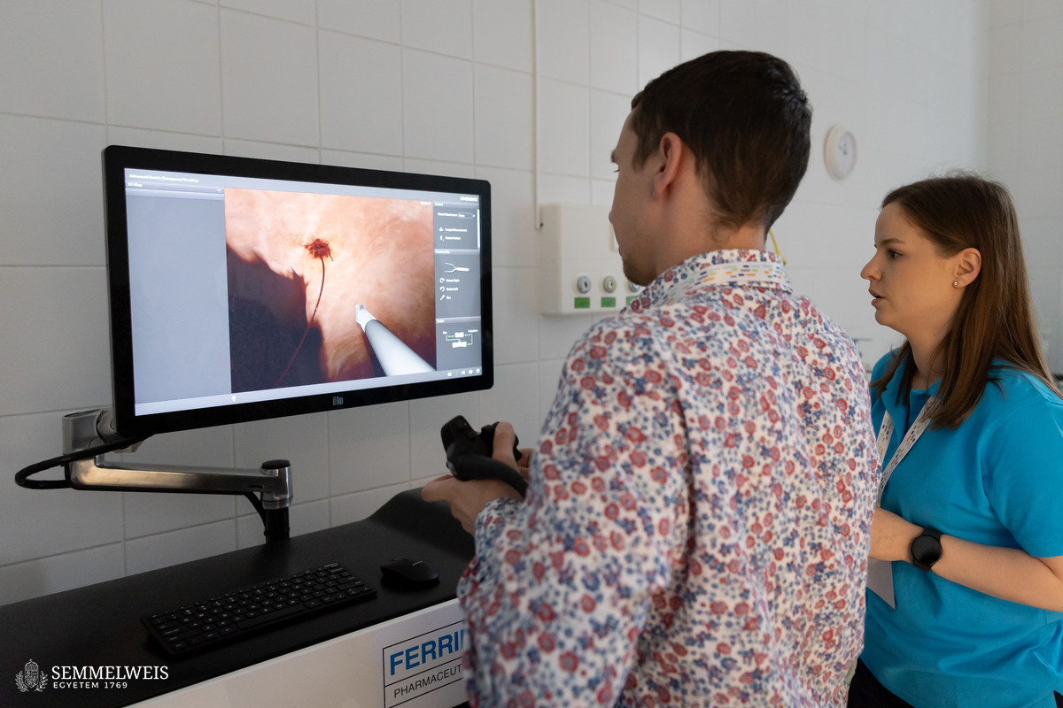
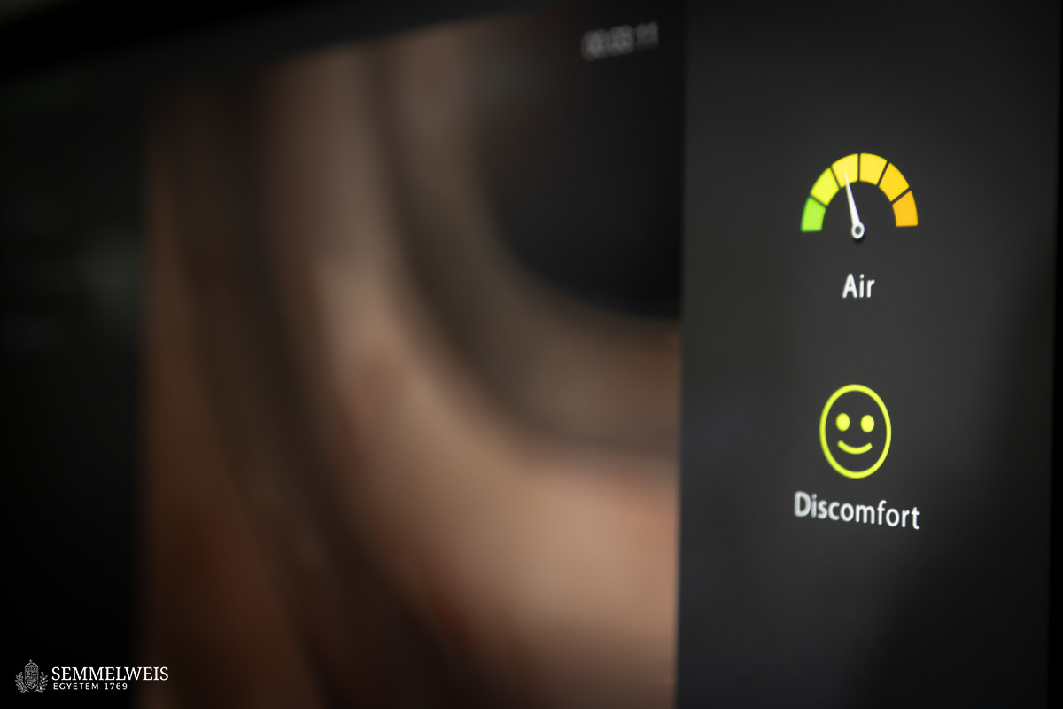
“To develop basic endoscopy skills, it would be enough to have a shoebox with a hole cut in it to attach various objects that could be searched and examined with the endoscope,” Dr. István Hritz remarked. The virtual simulator also features an introductory module where users can learn how to operate the device through various skill exercises. However, what makes the simulator special is that it recreates the situations encountered in endoscopic examinations in a realistic way. At the beginning of each exercise, the program outlines a medical case with the virtual patient’s data, medical history, and symptoms. During the endoscopic procedure, the trainee can monitor the object of examination and the movement of the device on the computer monitor. At the same time, the simulator provides audiovisual feedback on the progress of the procedure and the patient’s perception of pain. At the end of the exercise, the computer also evaluates performance.
Under the terms of the cooperation agreement between the university and the distributor, the simulator is to be initially used in the gastroenterology training for a period of one year; however, it will be available not only to Semmelweis students but also to domestic and international trainee specialists. The first such occasion was the international training course held at STéG on May 23-24, 2024, where participants had the opportunity to acquire these skills under the guidance of qualified simulator instructors Dr. Fruzsina Vilmos, trainee specialist, and Dr. Bálint Gellért, specialist in internal medicine.
Gallery
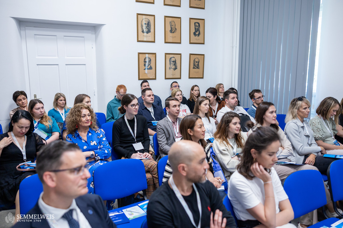
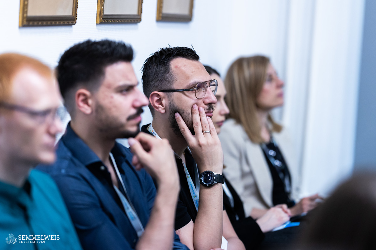
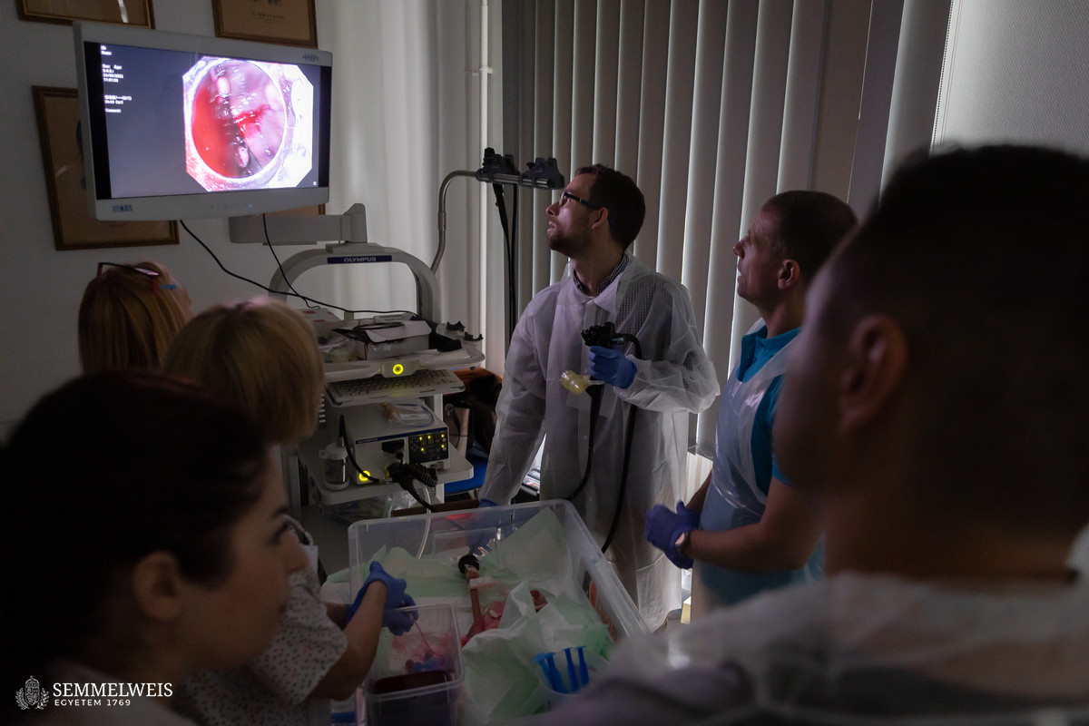
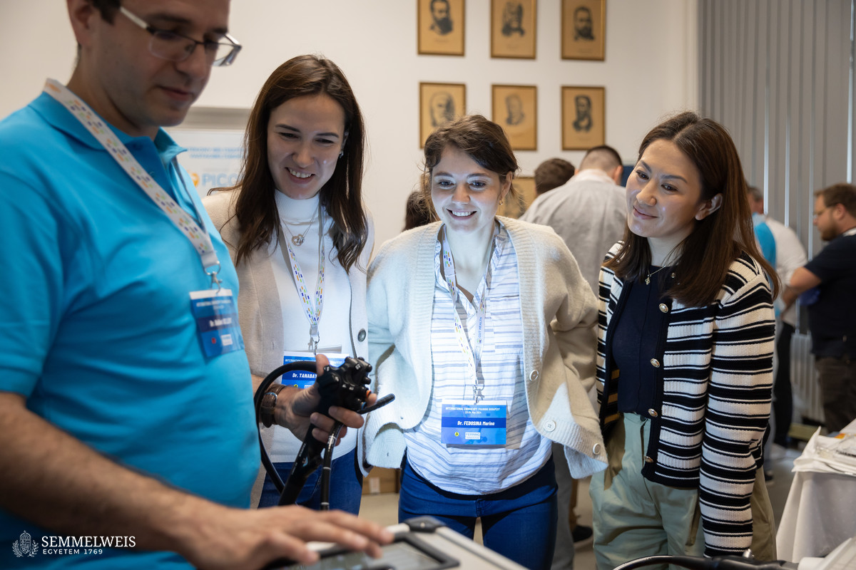
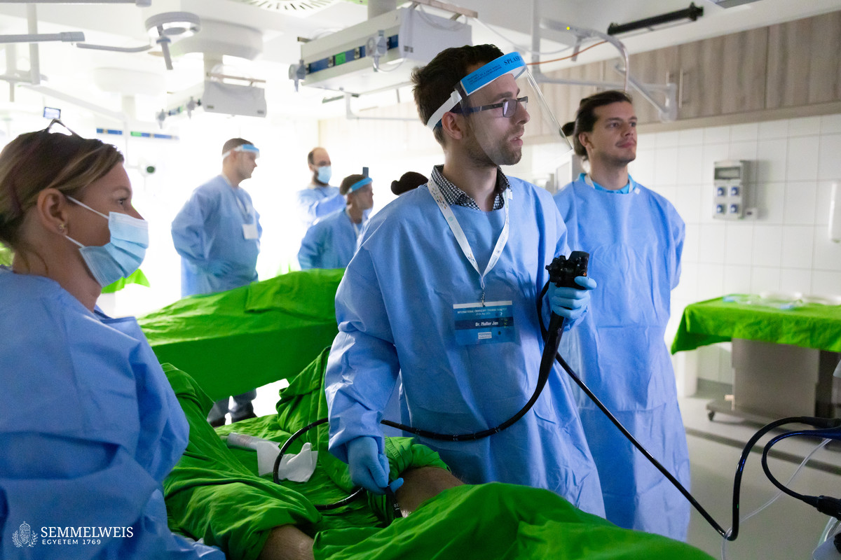
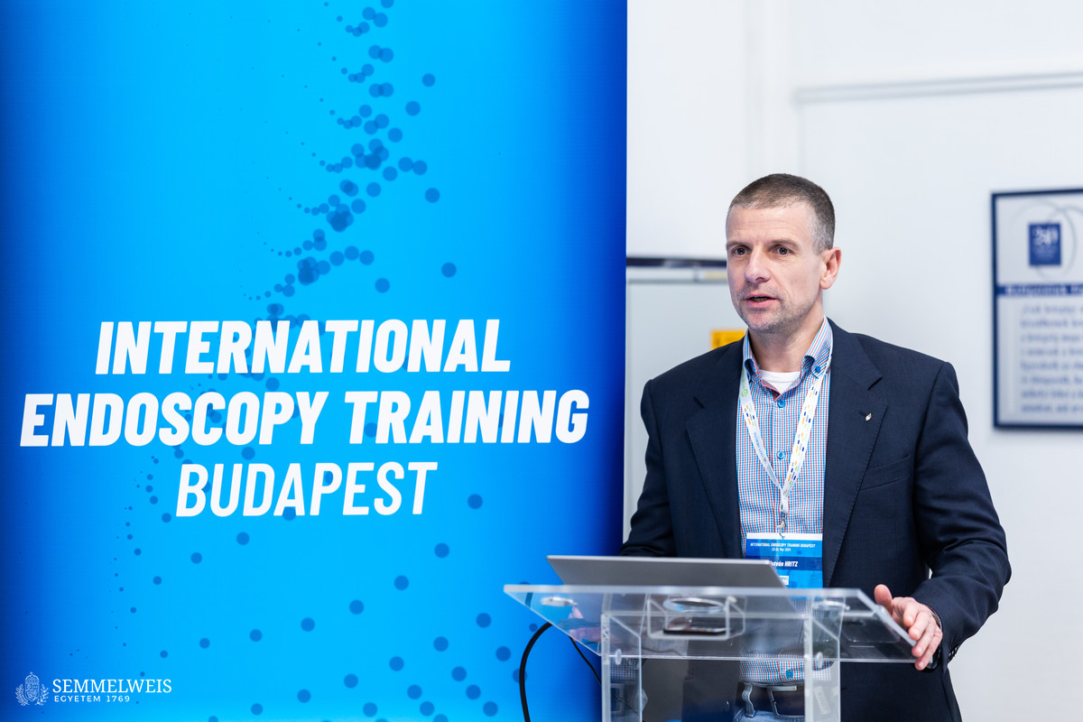
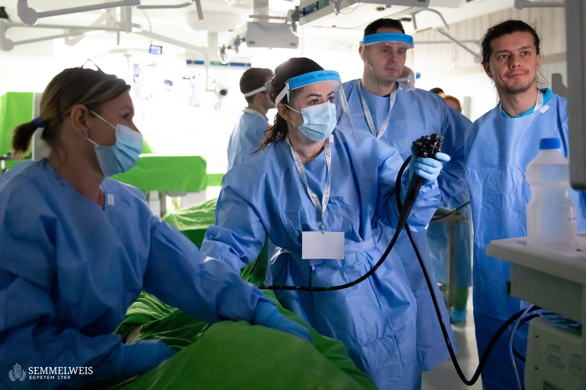
The International Endoscopy Training program was attended by twenty trainee specialists coming mainly from the region, Austria, Croatia, the Czech Republic, Romania, Serbia, Slovakia, and Kazakhstan, along with two Hungarian trainee specialists from STéG and the Department of Internal Medicine and Oncology. The participants had little or no prior experience with endoscopic interventions, so the first day of the two-day event was dedicated to theory, with seven presentations on various areas of endoscopy, delivered by international and Semmelweis University experts. The following day was devoted to five hands-on training sessions, each lasting 80 minutes, in rotating groups of up to five participants.
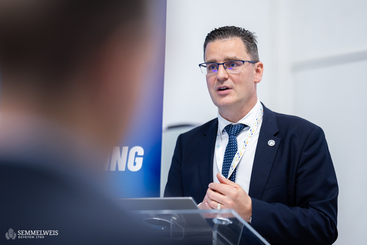
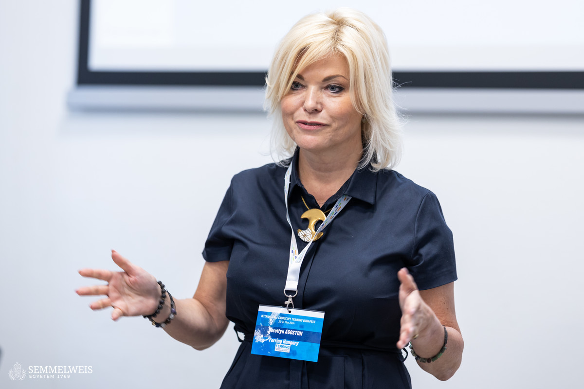
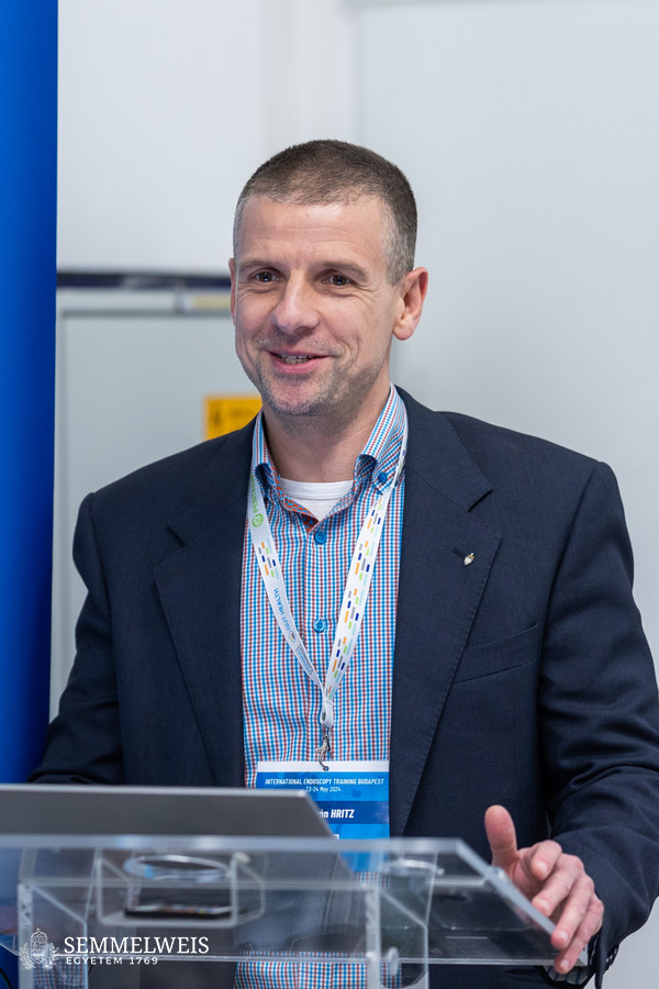
The event started with a short introduction and welcome by Dorottya Ágoston, Country Representative Hungary and Marketing Manager Gastro at Ferring Hungary Ltd., as well as speeches by Dr. Attila Szijártó, Director of STéG, and Dr. István Hritz, Head of the Invasive Endoscopy Unit of STéG and Chair of the Education Committee of the European Society of Gastrointestinal Endoscopy (ESGE).
Talking about the significance of the training, Dr. Attila Szijártó emphasized that participants would have the opportunity to learn hemostasis and perforation management techniques, gain insight into the cadaver endoscopy program, and master the basics of abdominal ultrasound examinations.
He also mentioned that their goal was to organize international training sessions twice a year and to take an active role in education in the country, for example through the education committee of the Hungarian Society of Gastroenterology (MGT).
 As Dr. István Hritz pointed out, what made the event special, in addition to the cadaver course, was the combination of the computer-based simulator with mechanical hybrid simulators, as well as with a composite porcine stomach. He also drew attention to the Endoscopy Museum at STéG, which he called unique worldwide as its collection includes some very old and special endoscopes as well as laparoscopic surgery tools; it also houses the first duodenoscope, with which the first ERCP (endoscopic retrograde cholangiopancreatography) – a test used to detect diseases of the bile ducts and pancreatic duct – was performed in Hungary in the early 1970s.
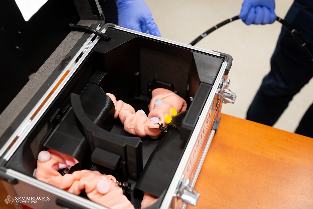
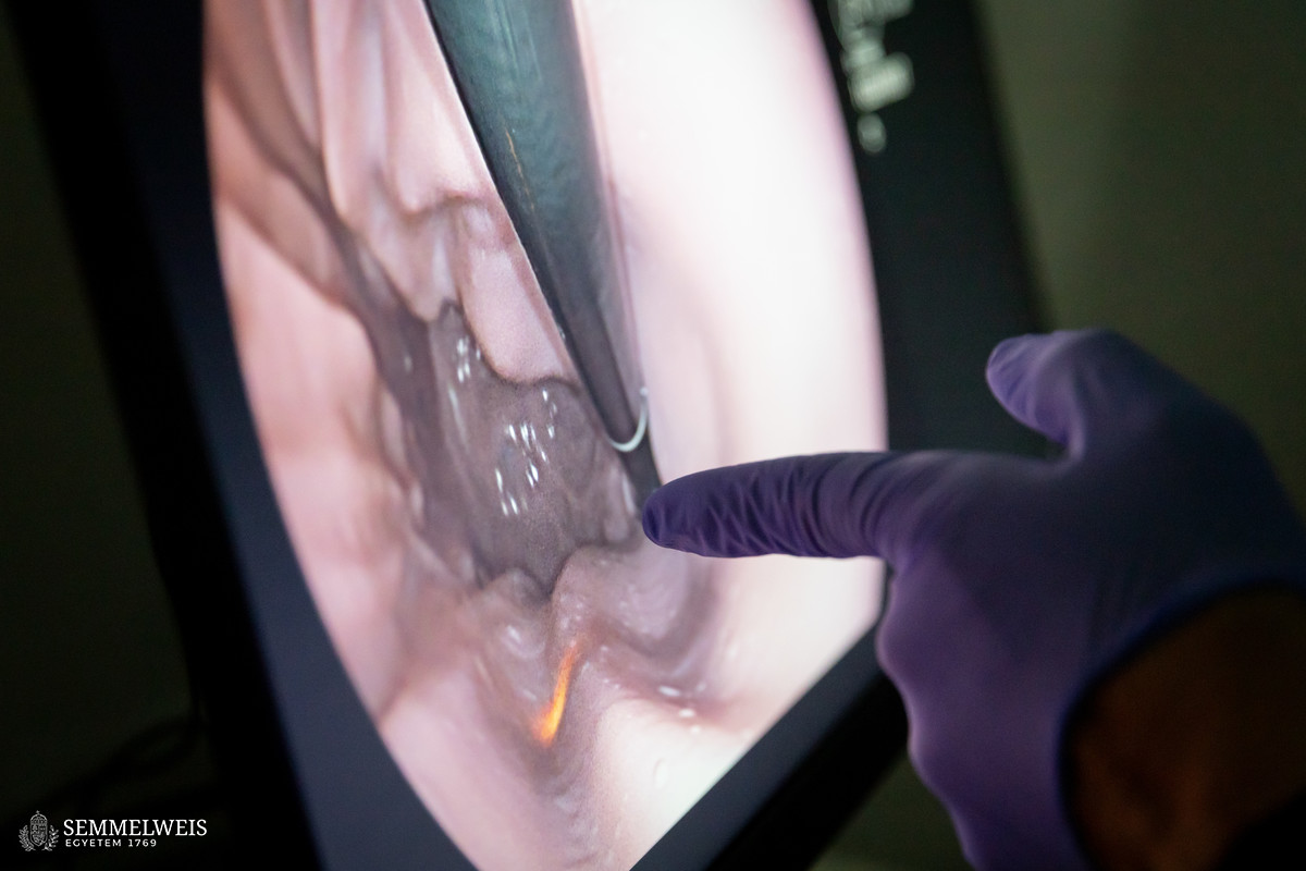
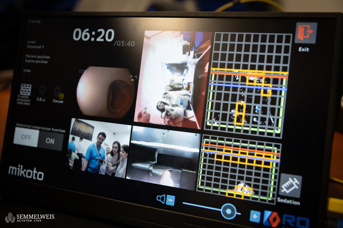
About the second day, which focused on hands-on training, Dr. István Hritz noted that for this occasion, the GI Mentor simulator, already available at STéG, was supplemented with endoscope towers, processors, and endoscopes used exclusively for training purposes, as well as with a Mikoto upper-lower tract hybrid simulator, provided by Hun-Med Ltd. and Endo Plus Service Ltd. Simulation with the latter device was supervised by Associate Professor Dr. Pál Miheller. The special feature of this simulator is that a latex stomach or intestine is attached to the computer hardware, which can sense how much pressure is applied or how -much air is pumped into the simulated organ by the trainee.
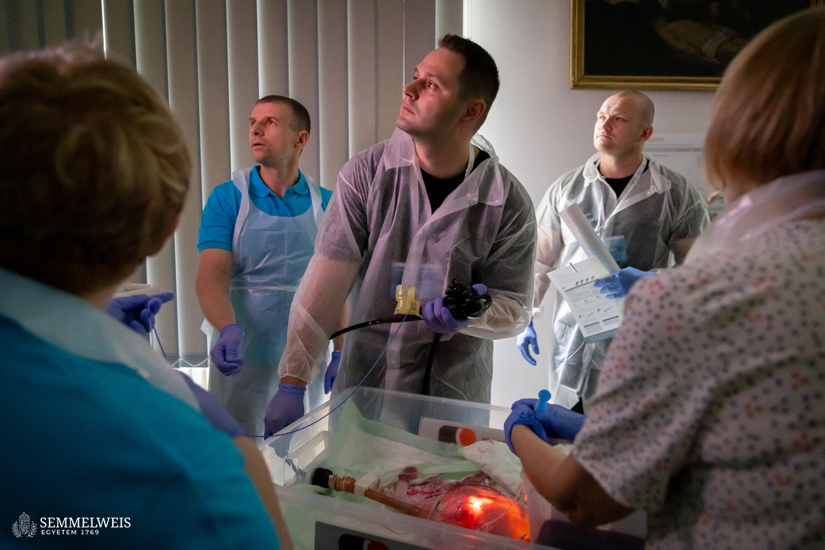
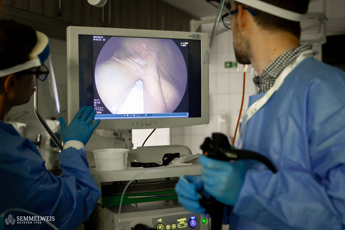
Another workshop, led by Dr. István Hritz, allowed participants to practice the management of hemostasis and perforation on porcine stomachs. The trainees placed ‘bear trap’ clips on simulated bleeding points using infusion catheters and beetroot juice. Another exercise modeled how perforation complications can be handled during an intervention.
 Also, in the framework of the two-day program but located in the Department of Anatomy, Histology and Embryology, trainee specialists had the chance to undergo cadaver endoscope training with the assistance of the staff at the Department of Internal Medicine and Oncology, Assistant Lecturer Dr. László Barkai and Assistant Professor Dr. Ákos Iliás. The fixation method attributed to Assistant Professor of Anatomy Dr. Tamás Ruttkay offers a unique training opportunity in the field of the stomach and intestines, which may even facilitate acquiring advanced endoscopy skills in the future.
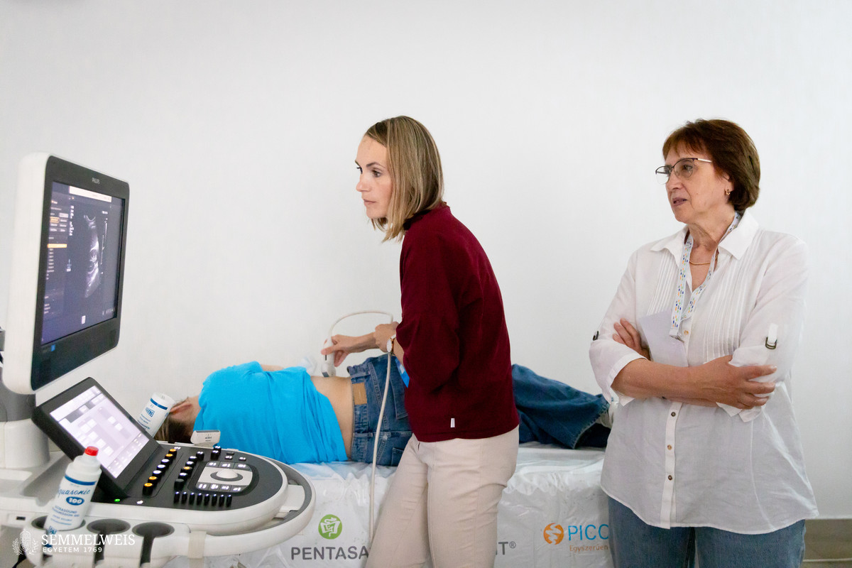
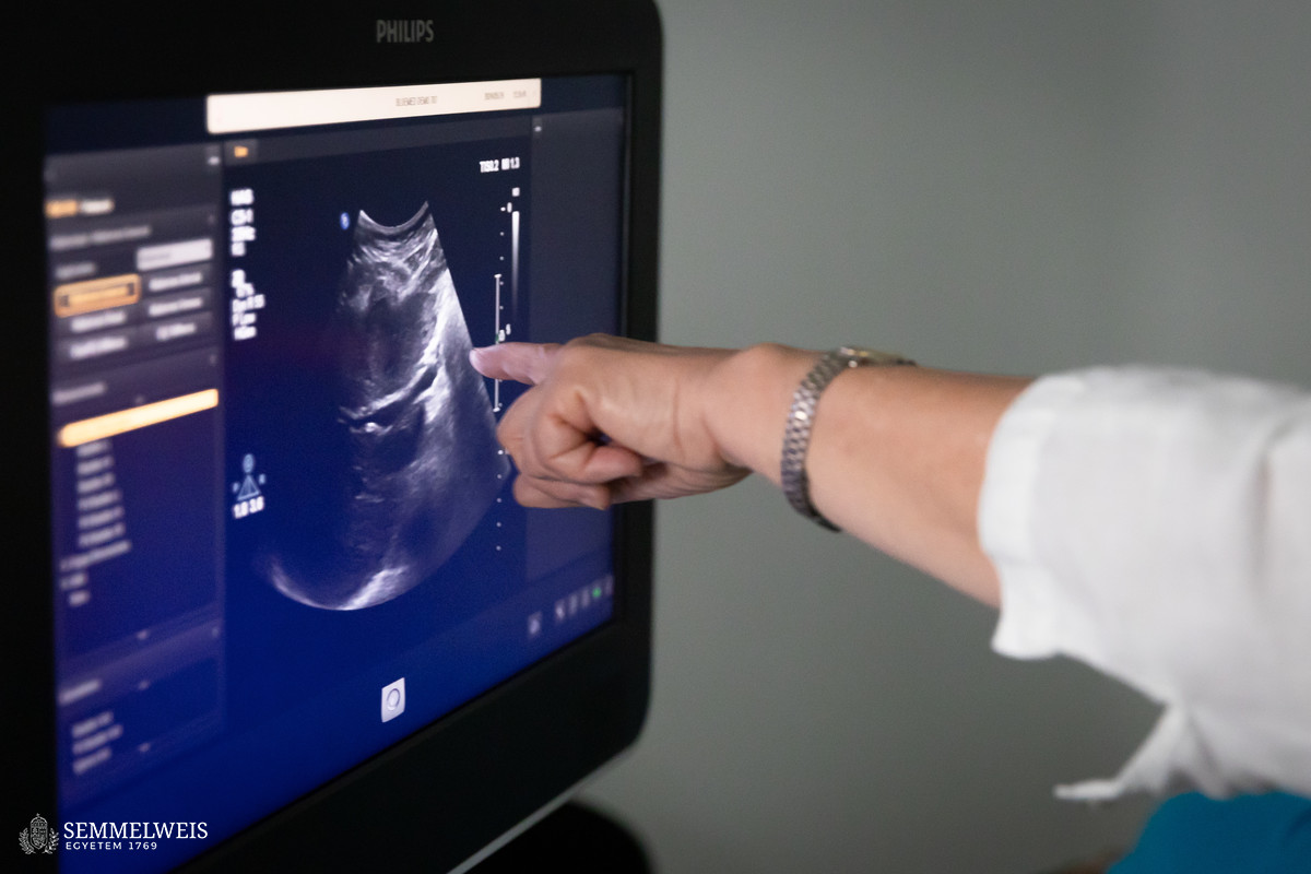
At the fifth workstation, under the professional guidance of Dr. Gabriella Győri, Consultant at the Medical Imaging Center, participants performed abdominal (liver, spleen, pancreas, kidney, and intestinal) ultrasound scans on healthy volunteer models. Although this is not an endoscopic examination, it is an essential part of the basic training of a gastroenterologist, stressed Dr. István Hritz.
The next international training course, also with the support of Ferring Hungary Ltd. and based on the experiences of the current two-day course, is scheduled for November 2024 at the Department of Surgery, Transplantation and Gastroenterology.
Dr. Balázs Csizmadia, Judit Szabados-Dőtsch
Photos by Bálint Barta, Boglárka Zellei – Semmelweis University