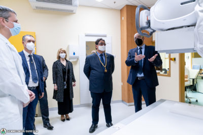
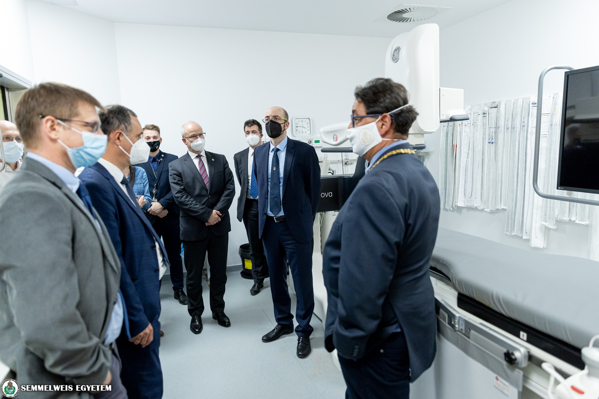
“During the last two years, the diagnostic imaging center has been completely transformed into a comprehensive medical imaging center under the leadership of dr. Pál Maurovich Horvat” – added rector dr. Béla Merkely during the visit. Dr. Merkely also added: Over the last few months several development projects have been introduced to enhance high-quality patient care. The visit took place on the National Day of Radiology, where dr. Merkely, dr. Pavlik and dr. Szabó inspected the brand new devices acquired within the development program in a value of approximately 600 million HUF. Dr. Pál Maurovich Horvat introduced the new generation equipments, including the mammography unit and the DSA laboratory, which is mainly specialized in oncological interventions.
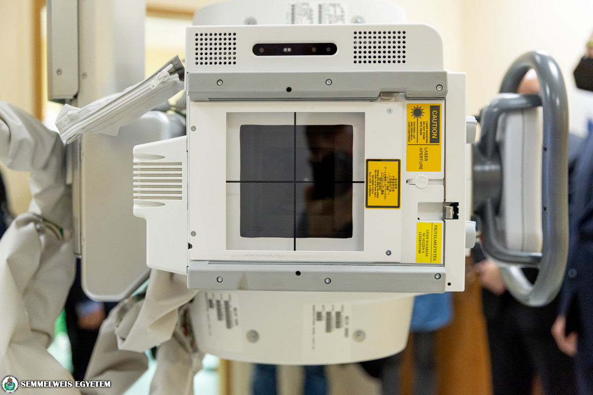
The two high-tech X-ray devices are currently only available here at Semmelweis University. The devices are able to detect 8 different types of pulmonary changes on chest X-rays using artificial intelligence.
Artificial intelligence supported medical imaging can enable a quicker and more precise clinical diagnosis and reduces the workload on radiologists.
– added dr. Pál Maurovich Horvat.
The new X-ray machines can assemble multiple images of the spine at high speed, allowing a thorough analysis of the entire spine. Furthermore, the device can be used for tomosynthesis, as it can create multiple three-dimensional, high definition images of the breast. In addition to traditional X-ray images, the equipment can also be used to produce dual energy images, allowing to effectively distinguish the bone structure from soft tissue – explained dr. János Gyebnár. The new X-ray machines are equipped with a special software called Thoracic Care Suite, which is able to automatically detect and identify a number of different pulmonary lesions, tumors, calcification, fibrosis and pleural effusion on chest X-rays. The Medical Imaging Center of Semmelweis University is the first European facility to apply this software in a clinical environment.
Gallery
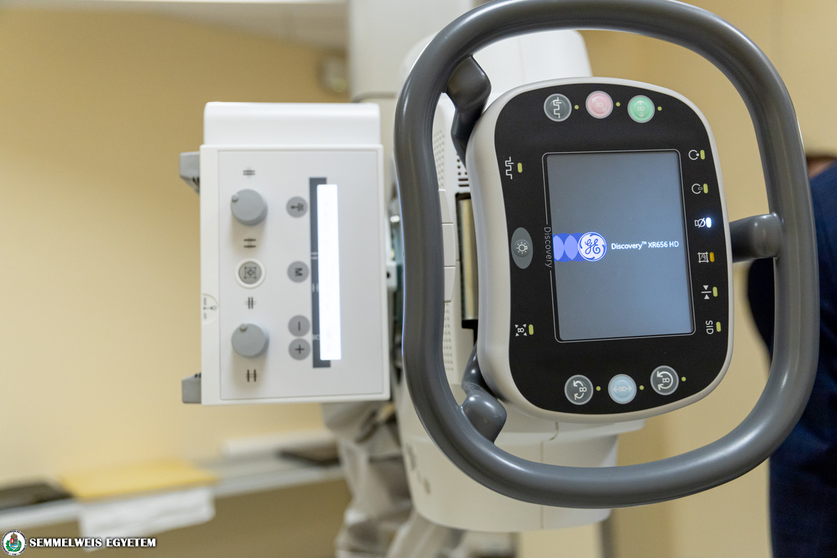
Artificial intelligence algorithms are designed to detect early signs of coronavirus, tuberculosis, and pneumonia. The new equipment enables to identify areas in the lung that may seem suspicious and are invisible to the bare eye. This way doctors may identify early-stage changes of a disease.
The other brand new software called “Critical Care Suite – Pneumotrax” can be used in emergencies and critical situations to identify signs of pneumothorax. The system examines the images by instantaneously searching for a pneumothorax. In case of a suspected disease, the AI algorithms evaluate the images and display the results on the monitor. This device is available to use in only a few healthcare institutions around the world.
The Medical Imaging Center acquired the two new X-ray devices (worth 200 million HUF) with the support of the Operational Staff and the Ministry of Finance.
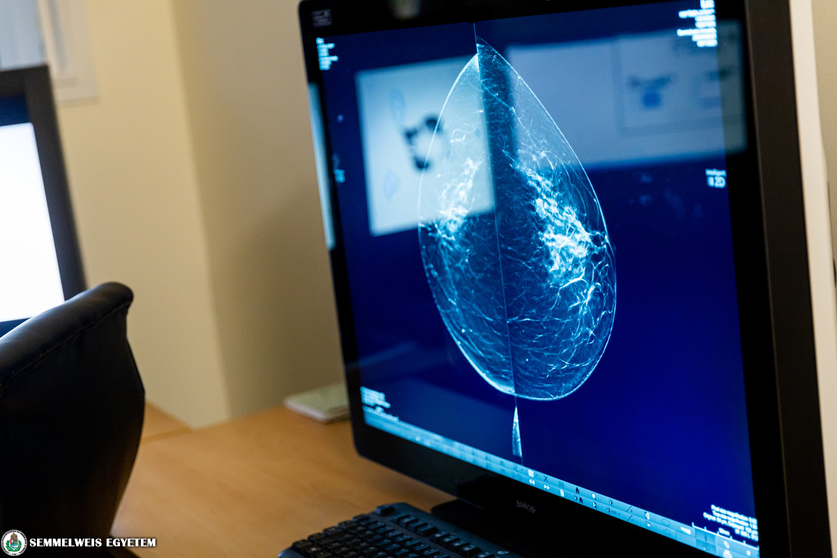
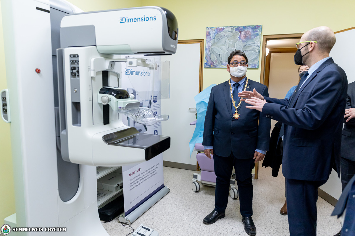
In September 2021, the Department of Breast Imaging of the Medical Imaging Center installed “Hologic 3Dimensions”, one of the most modern breast imaging devices in the world, which enables three dimensional tomosynthesis. This type of tomosytntheis is currently the most reliable and up-to-date breast imaging diagnostic system worldwide.
A major innovation introduced by this device is the bent compression plate, which mirrors the shape of a woman’s breast and allows both large and small breasts to be imaged in various exposures. The procedure involves less radiation exposure compared to the previous methods, X-ray images don’t get distorted and patients wont feel uncomfortable during the examination. During tomosynthesis, the X-ray tube continuously sweeps in a 15-degree arc over the breast to acquire a series of low-dose projection images at multiple angles. During a routine screening, the computer usually takes a maximum of 15 pictures of each breast at different angles (depending on the size of the breast), while during the conventional mammography procedure the specialized camera only takes two – pointed out dr. Mónika Tóth – chief clinician at the Department of Breast Imaging.
The tomosynthesis equipment uses the fastest and most modern technology available. With the help of the high definition 3D pictures taken by the machine, doctors can detect about 20-60% more breast cancer than using the traditional method. The approximately 100 million HUF worth device enables a much safer and quicker diagnosis in the same amount of time – added dr. Pál Maurovich Horvat.
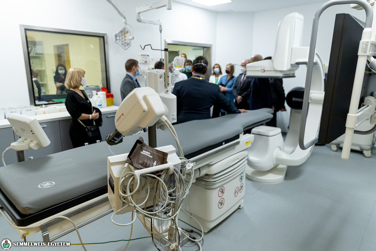
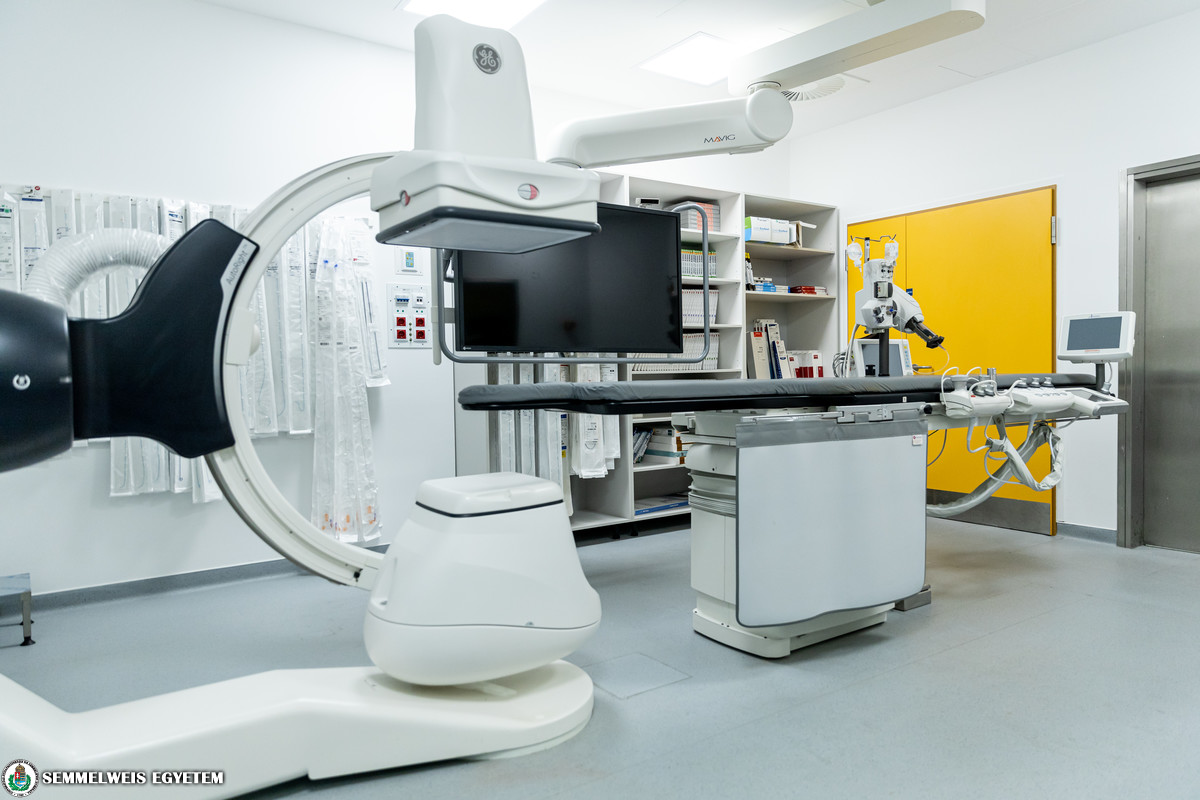
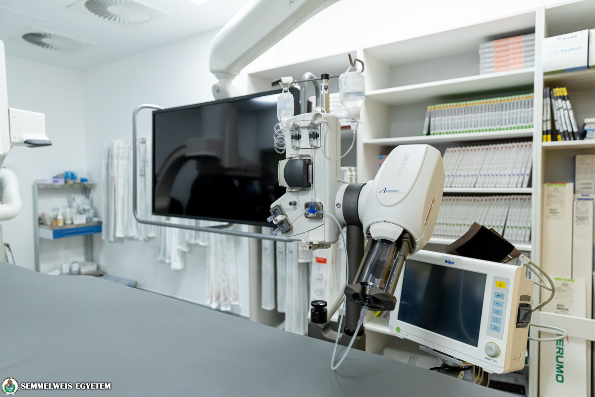
After just a couple of months operational, the new DSA laboratory enables doctors to perform high-quality oncological interventions.
“In this new environment, doctors will be able to perform major procedures, such as the embolization therapy for liver cancer, uterine artery embolization and prostatic artery embolization – added Balázs Nemes, head of the Department of Interventional Radiology. During the embolization procedure, an interventional radiologist usually inserts a catheter and guides it to the affected area. Then the doctor inserts small particles causing the vessels to become blocked. In case of cancerous diseases, medicine can also be injected this way.
Cutting edge IT technology at Semmelweis University
With the financial support of the Operative Board, the Medical Imaging Center of Semmelweis University has acquired a software package in the value of 80 million HUF. The software lets the staff of medical imaging facilities work as one across the network. The newest version of the Philips Concerto diagnostics work station is designed to enable a quicker and more reliable treatment of COVID-19 patients by connecting multiple clinical units of the university. The Philips AI software has been installed in line with the Medical Imaging Center’s innovative strategy.
Orsolya Dávid, Pálma Dobozi
Attila Kovács – Semmelweis University
Translation: Norbert Lukács