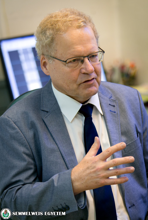
Around 40–50% of osteoporosis-related broken bone cases are preventable, which is why early detection and therapy are important, says Dr. Csaba Horváth, the head of the university’s Densitometry Laboratory, which performs the bone density scans that can diagnose the disease. The lab was established as the first of its kind in Hungary 35 years ago at the 1st Department of Internal Medicine, while the first bone densitometer in Hungary was also put into operation here, 40 years ago. Now the lab, which has held onto its position as a national leader ever since, is planning to follow international trends by developing densitometry’s function for measuring body composition.
 The densitometer that is used to diagnose osteoporosis by measuring bone density is a result of military developments during the Cold War: it owes its existence to the U.S. Apollo moon program that was launched as part of the Soviet-American space race, as astronauts are especially prone to osteoporosis because they lose bone density in zero gravity. With this machine, which was originally the size of a typewriter, they were able to continuously control the bone mineral content of those in space, says Dr. Csaba Horváth, a professor at the 1st Department of Internal Medicine and the head of the Densitometry Laboratory, providing some historical context.
He pointed out that the use of bone density measuring machines led to the discovery that osteoporosis is not a problem that only old ladies with bent backs have to contend with, but a widespread civilizational disease that affects one in every ten people. A visibly bent back is already the final stage; the bones start losing their density already years before, and can lead to broken bones even from a small trauma or fall. In Hungary, there are 100,000 such cases of broken bones, 40–50% of which are preventable, noted Dr. Csaba Horváth.
Part of the lost bone tissue can be replaced in quantity using available medications, but the damaged structure cannot, which is why early detection and therapy is important, the professor stressed. In Hungary, there are an estimated 300,000 people who have a level of osteoporosis that should be treated, while there are only 85,000 people currently receiving therapy nationwide, he said. The professor recommends that women get a bone density scan at the time of menopause, and then again two or three years later; in this way, the “fast bone losers” can be identified. In men, a scan should be done around age 50–60, but if there are some known risk factors (e.g. a history of the disease in the family, hyperthyroidism, liver or kidney diseases, medication that is harmful to bones, such as steroids), then it should be done regardless of age. An international tool for assessing risk of fracture due to osteoporosis (FRAX), which was developed over the past decade, has been in general use in Hungary as well; in fact, a workgroup of the Semmelweis department collaborated on the development of the tool, thanks to its decade-old partnership with the University of Sheffield. FRAX helps to evaluate the 10-year probability of bone fracture risk, based on densitometry results and other risk factors, and also provides guidance on whether pharmaceutical treatment should be started.
Addressing concerns over the side effects of the medication, Dr. Csaba Horváth said that the irritation problems affecting the mucous membranes of the stomach and esophagus can be avoided by taking medication using the right technique. For those patients that still do not tolerate the drug, it can also be administered by injection. Due to the dental side effects, professionals recommend that any necessary dental procedures be done prior to starting the medication. The professor also emphasized that osteonecrosis of the jaw occurs in only one out of every 20,000–30,000 patients, thus the benefits of the treatment far outweigh the risks.
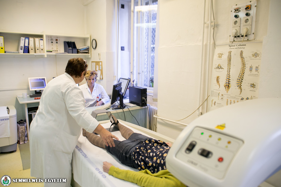
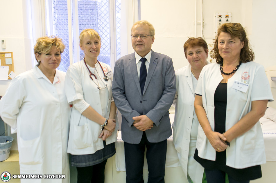 The 1st Department of Internal Medicine performs complex bone density scans on 3,500 patients a year. The examination, in which only a negligible amount of radiation is used, looks at the amount of X-ray photons that are absorbed by the body, indicating the calcium salt content of the bones. From the aspect of osteoporosis screening, the department qualifies as a national-level provider. In Hungary, this is where bone density measurement was developed and where it became an independent specialty; this laboratory introduced and developed its method of implementation in the country 30 years ago, highlighted Dr. Csaba Horváth, who also founded the lab. The first densitometer was brought to Hungary, to the 1st Department of Internal Medicine, around 40 years ago, in 1978, thanks to the department’s then-director Dr. István Holló. Today, bone density scans are performed at close to 100 locations in the country, which forms the basis for the national care of osteoporosis patients.
 The lab has retained its position as a national leader ever since: it acts as a collaborative partner in numerous studies regarding the bone-related aspects of certain diseases, it continuously plays a role in developing the densitometers, and one of its important areas of research is how the quality of bones can be measured, in addition to bone density quantity. The department’s Densitometry Laboratory also established the Osteology Working Group, which acts as a national cooperative professional forum and has organized free, credit point-providing further training courses for years. Additionally, they also organize national quality assurance testing and coordinate scientific studies.
Dr. Csaba Horváth also pointed out that in recent years, a completely new direction in the use of densitometry has started to evolve on an international level. Using a minimal amount of radiation, the devices can help determine body composition, i.e. in addition to bone mass, how much fat and muscle tissue can be found in the body. The quantity of the latter two is important in the case of obese, hyperlipidemic patients (who have high level of lipids, or fat) with high cardiovascular risk, as well as diabetics and gastroenterological disorders causing severe weight loss. The scan can also be used in sports medicine, for example to track the development of muscle mass. The measuring of body composition via densitometry is becoming increasingly widespread worldwide both in research and in treatment, therefore the laboratory plans to be a pioneer in developing this method of use in Hungary as well in the near future, emphasized Dr. Csaba Horváth.
Pálma Dobozi
Translation: Tamás Deme
Photo: Attila Kovács – Semmelweis University