In the retina, glycine release originates from glycinergic amacrine cell processes of the inner plexiform layer of the retina. This release of glycine may modulate retinal circuitry by activation of inhibitory glycine receptors and by acting as a coagonist on N-methyl-D-aspartate (NMDA) receptors both expressed on retinal ganglion cells [1]. The latter effect of glycine may be modulated by glycine transporter type 1 (GlyT-1) [2]. Cells with GlyT-1 immunopositive staining can be visualized in the inner nuclear layer and throughout the inner plexiform layer of the retina (Fig. 1.). Deprivation of oxygen and glucose (hypoxic conditions), which may occur in thromboembolic occlusion of retinal artery, induces excessive release of glycine (Fig. 1). In these conditions, GlyT-1 operates in the reverse mode and the resulted high levels of the NMDA receptor coagonist glycine released may be an additional factor in glutamate-mediated ganglionic cell damage. The GlyT-1 inhibitor NFPS (N-[3-(4’-fluorophenyl)-3-(4’-phenylphenoxy)propyl]sarcosine) does not alter the release of glycine from retina in normoxic conditions but inhibits glycine release during ischemia (Fig. 2). Thus, reduction of the NMDA receptor coagonist glycine concentrations in ischemic conditions by inhibition of GlyT-1 may decrease NMDA receptor-mediated neuronal toxicity and cell death in retinal tissue [3]. We conclude that GlyT-1 inhibitors may possess therapeutic value in treatment of the various neurodegenerative disorders of the retina such as glaucoma, diabetic or optic neuropathies, and retinal ischemia.
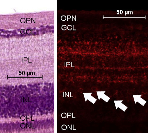 Fig. 1. Immunohistochemical study of chicken retina. Sections were stained with (A) hematoxylin-eosin (left side) and antiGlyT-1 (right side). The different layers of the chicken retina are indicated. Intensive staining of the GlyT-1 was found in the upper part of the inner nuclear layer and the inner plexiform layer. Visible cells stained with antiGlyT-1 are marked with arrows. OPN, optic nerve fibers; GCL, ganglion cell layer, IPL, inner plexiform layer, INL, inner nuclear layer; OPL, outer plexiform layer; ONL, outer nuclear layer.
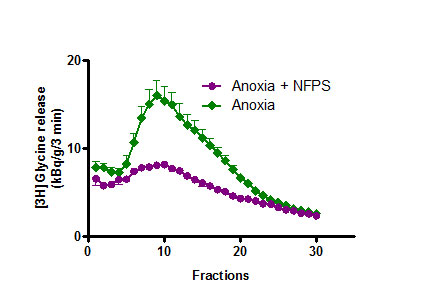 Fig. 2. Ischemia-induced [3H]glycine release from isolated chicken retina, this effect was reduced by addition of the GlyT-1 inhibitor NFPS (0.3 mM). Release of [3H]glycine by ischemic conditions (Krebs-bicarbonate buffer was glucose-free and saturated with 95% N2/5% CO2 gas mixture) was evoked from fractions 5 to 30. The GlyT-1 inhibitor drug was added to the retina at the beginning of fraction collection.
References:
[1]. Kertesz, Sz., Szabo, G., Udvari, Sz., Levay, G., Matyus, P., Harsing, L. G., Jr.,: Temporal alteration of spreading depression by the glycine transporter type-1 inhibitors NFPS and Org-24461 in the chicken retina. Brain Res., 2013, 1492, 1-6.
[2]. Harsing, L. G., Jr, Zsilla, G., Matyus, P., Nagy, K. M., Marko, B., Gyarmati, Zs., Szabo, A., Timar, J.:Interactions between glycine transporter type 1 (GlyT-1) and some inhibitor molecules. Acta Physiol. Hung., 2012, 99, 1-17.
[3]. Harsing, L. G. Jr., Albert, M., Matyus, P., Szenasi , G.: Inhibition of hypoxia-induced [3H]glycine release from chicken retina by the glycine transporter type-1 (GlyT-1) inhibitors NFPS and Org-24461. Exp. Eye Res., 2012, 94, 6-12.