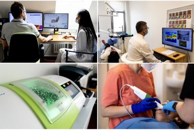
The Department of Restorative Dentistry and Endodontics is vigorously exploring several topics such as Digital Dentistry, Endodontics, Forensics, Oral Microcirculation, and Odontogenic infections.

Research zones

● Digital Forensic Dentistry

Members of the group: 
Principal investigator: Prof. Dr. János Vág 
Supervisor: Dr. Arvin Shahbazi 
Internal collaborator: Dr. Sándor Mikó
External collaborators: Prof. Dr. Katalin Nagy (Head of Oral Surgery Department, University of Szeged), Dr. Ajang Armin Farid (Head of the Hungarian Dental Unit in Disaster Victim Identification)
Ph.D. fellow: Dr. Botond Simon

Study title: Proof of concept of digital palatal morphology in human identification 

Abstract: The role of the rugae palatinae in forensic dentistry can be reinterpreted as a reliable method of human identification thanks to the development of digital dentistry. In our previous study, we have demonstrated the ability to distinguish monozygotic twins with almost identical DNA by comparing digitized samples of the palate. Our studies aim to assess the role of palatal geometry and surface morphology in the similarity between twins. By examining heritability, we will determine the role of genes and environment in the variation between individuals. We want to determine the extent of palate degradation after death under different environmental conditions and how long the characteristics required for identification can be recognized. We aim to demonstrate the stability of palatal tissue by periodontal plastic surgery post-operative regeneration examination and histological studies. Our expected results suggest that palatal morphology is resistant to degradation for 1-2 weeks and thus could be well suited for use in victim identification. We aim to demonstrate that differences in software and hardware between intraoral scanners used by dentists do not affect identification accuracy. This will allow comparisons between antemortem (reference database) and future postmortem samples using intraoral scanners. Based on the results of our studies, a pattern recognition algorithm supported by machine learning can be developed. Our evidence-based results may trigger the international exploitation of digital palatal identification.

Endodontics

Members of the group:

Principal investigators: Dr. Enikő Szabó, Dr. Zsolt Lohinai 
Internal collaborator: Dr. Brigitta Huszta, Dr. Melinda Polyák, 
Postgraduate fellows: Dr. Dániel Cifrák 

Study title: Effectiveness of endodontic irrigants 

Abstract: In secondary/persistent endodontic infections, disinfection of the root canal is a challenge for the endodontist. There is a need for irrigants that can penetrate into the mechanically unreachable areas of the canal. The limitation of sodium hypochlorite is its significant surface tension, which may result in restricted penetration of the irrigant into small tubules and canals. 

Effectiveness of endodontic irrigants in teeth with chronic periapical lesions 
In our study, we compare the effectiveness of sodium hypochlorite and hyper-pure chlorine dioxide. After retreatment, the tooth with periapical lesion will be followed for four years clinically and radiologically. During root canal treatment, microbial samples are taken from the canal: 1) from the infected canal after removing the root canal obturation, 2) after disinfection, and a one-week temporization. The microbial composition of the samples are evaluated qualitatively and semi-quantitatively by culturing and molecular methods.

Effectiveness of endodontic irrigants in the depth of dentin along the tubules 
The study is performed on the distal root of lower molars. After amputating the root, it is mechanically prepared and sterilized in an Eppendorf tube. It is then artificially contaminated by a test bacteria, Enterococcus faecalis. The root is treated by two disinfectant irrigants. After splitting and staining with fluorescent viability stain, the functional penetration of the disinfectants along the tubules is evaluated by confocal laser scanning microscopy.

Prevalence of parodonthopathogens in the pulp and periodontal pocket of teeth with periodontitis 
Marginal periodontal inflammation may spread to the pulp and the other healthy pulpal tissues. In our study, we take microbial samples from the periodontal pocket and the pulp of coronally intact teeth extracted because of severe periodontitis. We determine the prevalence of 11 parodontopathogens in the pulp. We look for a correlation between the severity of periodontitis and the status of the pulp. The presence of inflammatory and necrotic factors are determined by molecular methods.

● Microcirculation 

Members of the group:
Principal investigator: Prof. Dr. János Vág 

Supervisors: Dr. Réka Fazekas, Dr. Eszter Molnár, Dr. Bernadett Gánti 
External collaborator: Dr. Szabolcs Várbíró
Ph.D. fellow: Dr. Barbara Mikecs 

Study title: Investigation of the regulation of the gingival microcirculation 

Abstract: Our study party, in the past seven years, performed many physiological tests by investigating the gingival microcirculation, from which publications have appeared. After applying the post occlusive reactive hyperemia test (PORH), we could monitor the spatial and temporal changes in the blood flow of the human gingiva. During the short-term (5 second), compression in the marginal gingiva ischemia was observed, which was followed by 5 minutes of lasting hyperemia. This was the horizontal PORH test. Both ischemia and hyperemia extended to areas further away from the compression. In men, this was greater and more prolonged than in women, and in men, we observed a „rebound” effect, which in women was not present. Our study group has successfully applied the PORH test to compare the blood flow of the gingival tissues surrounding natural teeth and dental implants. In other physiological tests, we’ve investigated the effect of different agonist solutions on gingival microcirculation. In recent years we could prove the presence of spreading vasodilation and spreading vasoconstriction in the human gingiva. Currently, we investigate the endothelium-dependent and endothelium nondependent vasodilation after applying Nitric oxide (NO) or acetylcholine donor solutions. We are looking for an explanation regarding the function of the endothelium and collaterals of the attached gingiva between males and females. By applying the PORH test and donor solutions, currently, we are investigating the impact of age and female sex hormones on gingival microcirculation. These physiological investigations may shed light on gender differences depending on hormonal and age-related effects and may provide information on the functioning of microcirculation under physiological conditions. These data may serve as a control for further studies in pathological conditions that also significantly affect microcirculation, such as diabetes or periodontal disease.

● Microcirculation 

Members of the group:
Principal investigator: Dr. Eszter Molnár  
Specialist: Dr. Péter Komora 
Specialist candidate: Dr. Roland Daubner  

Study title: The role of pulp microcirculation in the diagnosis and treatment of pulp diseases 

Abstract: A new trend has appeared in the classification of pulpitis in the international literature. These classifications also influenced the consequent therapeutic care, emphasizing various vital pulp therapies rather than the previously preferred lege artis root canal treatment. However, no precise recommendation has been made for heterogeneous pulp preservation treatments such as pulp capping or pulpotomy. Furthermore, it is not always easy to make a correct diagnosis, as we are not able to perform a histological examination in case of pulpitis. Laser Doppler blood flow measurement is a widely used non-invasive real-time method in studying microcirculation. This technique is also suitable for measuring dental vitality in addition to local cold and electrical tests, which are widely used to determine sensitivity. So we can obtain data on the blood flow of the pulp in addition to its neural responsiveness. In our study, we would like to determine whether we can make a diagnosis of the current condition of the dental pulp in a way that accurately determines therapy and whether we can correlate the diagnostic classification with blood flow measurement. Respectively, we are looking for the answer to whether the laser Doppler flowmeter can be helpful as a diagnostic tool or during treatment. A pain scale describing the patient’s complaint and additional radiological examinations are also used to establish the diagnosis. Therapeutic care is performed according to current ESE recommendations using a dental operating microscope, and study participants are followed after endodontic care, examining the rate of successful interventions. Our evidence-based results may help in the day-to-day, accurate application of the therapies suggested by the new endodontic classifications.

● Microcirculation 

Members of the group:
Principal investigator: Dr. Réka Fazekas
Internal collaborator: Dr. Eszter Molnár
External collaborators: Dr. Bálint Molnár (Semmelweis University, Department of Periodontology), Dr. László Kőhidai (Semmelweis University, Department of Genetics, Cell- and Immunobiology), Dr. Ferenc Bartha (Semmelweis University, Department of Periodontology), Dr. Afrodité Kőrösi (Semmelweis University, Department of Periodontology), Dr. Orsolya Láng (Semmelweis University, Department of Genetics, Cell- and Immunobiology)
Ph.D. fellow: Dr. Eleonóra Sólyom

Winning application: OTKA FK_2020 – 135348 (topic leader: Dr. Bálint Molnár)

 
Study title: Blood flow kinetics following human alveolar ridge augmentation procedure measured by Laser Speckle Contrast Imager
 
Abstract: In periodontal surgery, various incisions, flaps, grafts, and wound closure techniques are used, resulting in varying degrees and durations of ischemia. However, postoperative wound healing is significantly influenced by the preservation of the gingival microvasculature and the revascularisation of the surgical site. Our working group aims to study the effects of different types of surgery on regeneration. In our studies, we use a Laser Speckle Contrast Imager (LSCI), which can be used non-invasively and reproducibly to measure capillary blood flow, even covering the entire surgical site, thus providing an objective comparative assessment of the impact of surgical factors on healing. Previously, we successfully used LSCI to characterize the reperfusion curves of regions distinguished in the surgical field while incorporating various grafts and individually monitoring oral wound healing. In addition to blood flow measurements, we complement our studies on wound healing by studying angiogenesis, i.e., the quantification of wound fluid and measuring Vascular Endothelial Growth Factor (VEGF) expression.


● Odontogenic infections and related treatments

Members of the group:
Principal investigator: Dr. Zsolt Lohinai  
External collaborator: Dr. Martin Levine (Department of Biochemistry, Colleges of Medicine and Dentistry, University of Oklahoma)
Ph.D. fellows: Dr. Kasidid Ruksakiet, Dr. Mikaela Aresti 

Study titles:

    ● New Concepts Concerning Lysine Metabolism in Periodontal Disease. 

    ● Antimicrobial efficacy of chlorhexidine and sodium hypochlorite in root canal disinfection.

Abstracts: Bacterial lysine decarboxylase influences human dental biofilm lysine content, biofilm accumulation, and subclinical gingival inflammation Background: Dental biofilms contain a protein that inhibits mammalian cell growth possibly lysine decarboxylase from Eikenella corrodens. This enzyme decarboxylates lysine, an essential amino acid for dentally attached cell turnover in gingival sulci. Lysine depletion may stop this turnover, impairing the barrier to bacterial compounds. This study aims to determine biofilm lysine and cadaverine contents before oral hygiene restriction (OHR) and their association with plaque index (PI) and gingival crevicular fluid (GCF) after OHR for one week. Methods: Laser-induced fluorescence after capillary electrophoresis was used to determine lysine and cadaverine contents in dental biofilm, tongue biofilm, saliva before OHR, and dental biofilm after OHR. Results: Before OHR, lysine and cadaverine contents of dental biofilm were similar and 10-fold greater than in saliva or tongue biofilm. After one week of OHR, the biofilm content of cadaverine increased, and that of lysine decreased, consistent with greater biofilm lysine decarboxylase activity. Regression indicated that PI and GCF exudation were positively related to biofilm lysine after OHR, unless biofilm lysine exceeded the minimal blood plasma content, in which case PI was further increased, but GCF exudation was reduced. Conclusions: After OHR, lysine decarboxylase activity seems to determine biofilm lysine content and biofilm accumulation. When biofilm lysine exceeds minimal blood plasma content after OHR, less GCF appears despite more biofilm. Lysine appears essential for biofilm accumulation and the epithelial barrier to bacterial pro-inflammatory agents. Inhibiting lysine decarboxylase may retard the increased GCF exudation required for microbial development and gingivitis. 

Chlorine dioxide and halitosis 
sssssss
Members of the group:
Principal investigator: Dr. Beáta Kerémi 
Ph.D. fellow: Dr. Eszter Szalai
Project students: Saghar Shojazadeh, Viktória Babay, Adél Galvács
sssss
Study title: Chlorine dioxide and halitosis 
sssss
Abstract:
Effectiveness of chlorine dioxide
Our group investigates the effect of chlorine dioxide mouthwashes in various conditions like periodontitis and halitosis with meta-analyses. We aim to prove the effectiveness of chlorine dioxide and give clear guidance on which mouthwashes should use. We can change the routinely used mouth rinses, which are full of side effects. We want to suggest an evidence-based treatment protocol for halitosis.
ssss
Measurement of halitosis
We can use the gold standard organoleptic measurement to measure halitosis, which is a subjective method. Another option is to use various objective devices, for example, OralChroma, halimeter, and eNose. All methods have advantages and disadvantages, but we aim to find the best one to measure halitosis and suggest it to colleagues. We hope it can standardize the diagnosis and helps to offer also individual therapy for patients.
sssss
Randomized clinical trial in halitosis

Well-treated halitosis can improve patients’ well-being; we plan a randomized clinical trial to compare more types of mouthwashes in patients with bad breath. We will compare the most used mouthwashes and chlorine dioxide, which lack known side effects, and not just eliminate the bacteria that produce volatile sulfur compounds from sulfur-containing amino acids that cause bad breath and also decrease the amount of unpleasant smelling volatiles.