CardioLab is the cardiac CT research and core laboratory of the Heart and Vascular Center, Semmelweis University, Budapest. We connect a high-volume clinical cardiac CT program with quantitative imaging research, so that advances in CT translate into better risk assessment, treatment decisions and patient care.
2
CT systems: photon-counting CT (NAEOTOM Alpha.Peak) and dual-source CT (SOMATOM Force)
4,500+
cardiac CT examinations per year
Level III
EACVI-certified cardiac CT experts lead the program
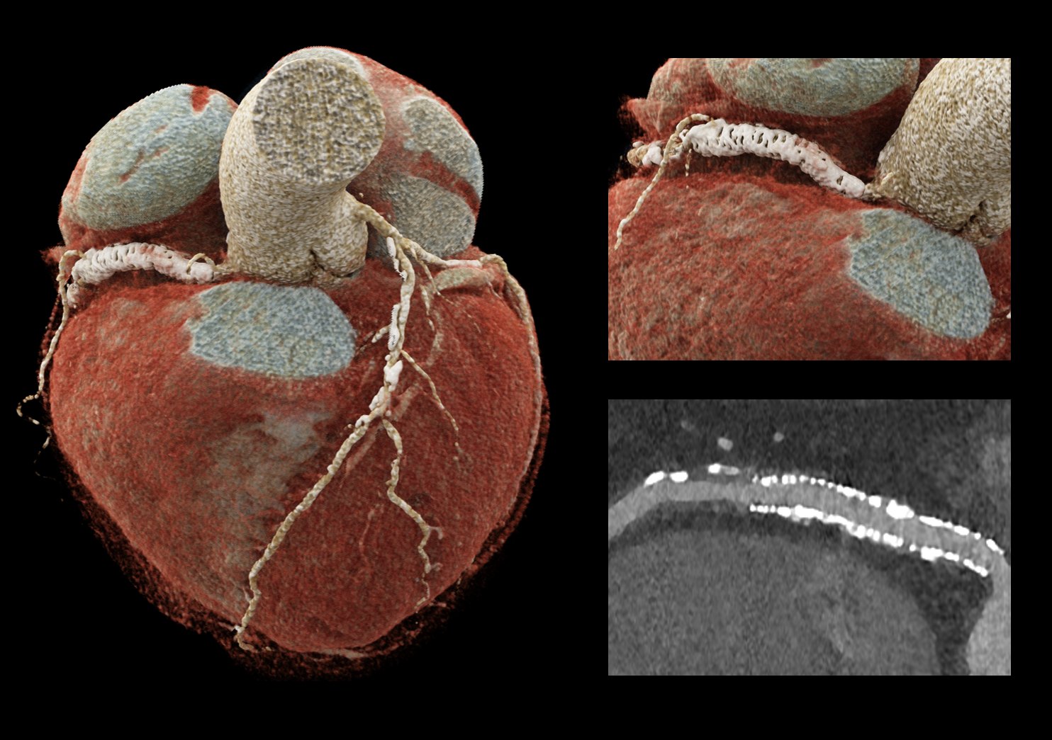
Coronary CT angiography: volume rendering and curved planar reformation of a calcified right coronary artery.
What we do
Clinical imaging, research and core lab under one roof
Clinical cardiac CT
All aspects of cardiac imaging for the Heart and Vascular Center: coronary CT angiography, structural and procedural planning, aortic imaging and post-transplant follow-up.
Research
Quantitative plaque and pericoronary adipose tissue phenotyping, myocardial perfusion, photon-counting CT, novel post-processing tools, outcome studies and AI-based approaches for novel biomarkers.
Core laboratory
Standardised CT analysis for sponsored and investigator-initiated multicentre trials, including longitudinal plaque progression studies and CT-guided PCI.
Our focus is clinical translation: we evaluate how new CT technology and analysis tools change diagnosis, risk stratification and management, not only image quality. We work in close collaboration with the electrophysiology and haemodynamic laboratories and with the other cardiac imaging modalities of the Heart and Vascular Center.
Our approach to AI
Independent clinical validation
CardioLab does not develop its own AI software. We clinically validate third-party, AI-based plaque analysis and CT post-processing tools against established reference standards, providing independent evidence on their accuracy and clinical value. Together with partner research groups, we also test new technologies — for example, the capabilities of photon-counting CT detectors in dedicated phantom studies.
Experience
Ten years of cardiac CT: how our expertise was built
Our expertise has grown step by step since 2015: from image acquisition and reconstruction, through quantitative plaque and functional imaging, to photon-counting CT and outcome research. Each step was built on systematic validation against reference standards.
2015–2017
Image quality, reconstruction and protocols
We started with the fundamentals of CT acquisition and image reconstruction, including how iterative model reconstruction changes coronary artery calcium scores. This work laid the technical basis for reliable quantitative measurements.
Key paper: Int J Cardiovasc Imaging 2015
2017–2020
Quantitative plaque, function and structural imaging
We moved from image quality to quantitative phenotyping: structured CAD-RADS reporting, CT-derived left ventricular and left atrial strain validated against echocardiography, and subclinical leaflet thrombosis and reverse remodelling after TAVI.
Key papers: J Cardiovasc Comput Tomogr 2017 · J Cardiovasc Comput Tomogr 2019 · Eur Heart J Cardiovasc Imaging 2020
2020–2023
Plaque progression and perfusion
Serial CT showed that CAD-RADS can miss plaque progression in almost half of patients, which led to our practical guidance on how to quantify coronary plaque. We also linked total plaque volume to ischaemia on dynamic perfusion CT.
Key papers: Eur Heart J Cardiovasc Imaging 2022 · Eur Heart J Cardiovasc Imaging 2022 · Front Cardiovasc Med 2022
2023–today
Photon-counting CT and clinical translation
With photon-counting CT we systematically compared ultra-high-resolution imaging with energy-integrating CT: stenosis against quantitative coronary angiography, plaque quantification and in-stent restenosis. In parallel, outcome analyses showed that the benefit of statins increases with CT-defined disease severity, and that high-risk plaque carries prognostic weight comparable to obstructive disease.
Key papers: Eur Radiol 2023 · Circ Cardiovasc Imaging 2024 · Radiology 2025 · JACC Cardiovasc Imaging 2025 · Eur J Radiol 2026 · Eur Radiol 2026
Validation track record
What we have validated
Reconstruction algorithms
Iterative, model-based, quantum iterative and deep-learning reconstruction: effects on calcium score, plaque volumes, image noise and stenosis measurement.
CT strain
Left ventricular and left atrial strain by CT against echocardiography; atrial strain and reverse remodelling after TAVI.
Plaque analysis software
Semi-automated and AI-based plaque quantification tools: reproducibility and agreement across energy-integrating and photon-counting CT.
CT-FFR
On-site CT-FFR against invasive FFR, including non-culprit lesions after acute coronary syndrome.
Photon-counting CT vs invasive reference
Stenosis against quantitative coronary angiography, obstructive disease against invasive angiography, and in-stent restenosis, including OCT-confirmed cases.
Phantom ground truth
Calcium, iodine and low-attenuation plaque fidelity in dedicated phantoms, with optimisation of kernels, reconstruction strength and energy levels.
Current focus
Where ten years of validation work is heading now
Plaque validation on photon-counting CT
Phantom and intravascular imaging (IVUS/OCT) as ground truth for plaque quantification.
Intervention planning
CT-guided PCI, calcified lesions, stents and in-stent restenosis.
Perfusion
Dynamic myocardial CT perfusion and tissue characterisation with photon-counting CT.
Leadership
Who leads CardioLab
The lab is led by Bálint Szilveszter, MD, PhD, FEACVI, FSCCT, Assistant Professor of Cardiology and head of the Cardiac CT Working Group at the Heart and Vascular Center.