Cardiac CT Core Lab & Research · Semmelweis University
Cardiovascular CT from phenotype to patient care
Cardio Lab is a clinically integrated cardiovascular CT research and core laboratory platform at Semmelweis University. Our work spans the cardiovascular care pathway from disease detection and prevention to deep phenotyping, functional assessment, treatment monitoring, and image-guided intervention.
We combine quantitative cardiovascular imaging, clinical validation of AI-based analysis tools, photon-counting CT, myocardial perfusion, longitudinal drug trials, phantom research, and procedural planning to determine how advanced CT can improve risk assessment, therapeutic decisions, and patient care.
4,500+
cardiac CT examinations per year
PCCT
NAEOTOM Alpha.Peak photon-counting CT in clinical routine
Level III
EACVI-certified cardiac CT experts
Our approach
From imaging technology to patient care
We do not study single biomarkers or technologies in isolation. Cardio Lab covers the full translation pathway — acquisition, quantification, AI, validation, phenotyping, prevention, therapy, intervention and outcomes — so that advances in CT reach clinical decisions. We work in close collaboration with the electrophysiology and haemodynamic laboratories and with the other cardiac imaging modalities of the Heart and Vascular Center.
Prevent →
Phenotype →
Understand →
Treat →
Intervene →
Improve care
Core Lab & AI
AI-based quantitative imaging and clinical validation
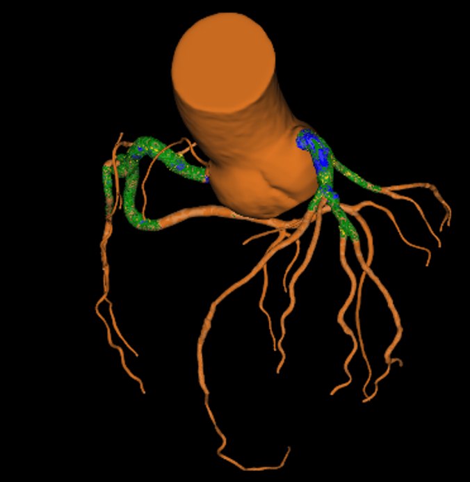
From automated measurements to clinically validated biomarkers. Cardio Lab does not develop its own AI. We provide an independent validation environment for third-party, AI-based plaque analysis and CT post-processing tools — testing accuracy against reference standards and asking whether the numbers change risk assessment and management.
Our core lab performs standardised quantitative analyses for sponsored and investigator-initiated multicentre studies, built on a high-volume clinical program.
Core Lab capabilities
- Coronary plaque quantification: total, calcified, non-calcified and low-attenuation plaque
- High-risk plaque features and CAD-RADS stenosis grading
- Pericoronary adipose tissue (PCAT) attenuation
- Serial CT analysis for drug trials
- Independent validation of AI-based analysis tools
- Protocol and image-quality support for participating sites
Prevention
Prevention, reporting and adherence
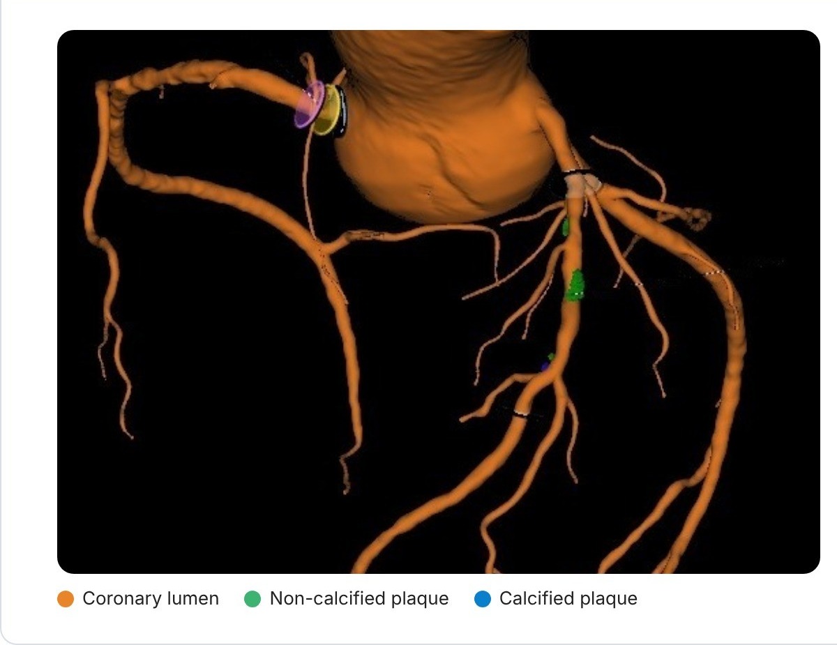
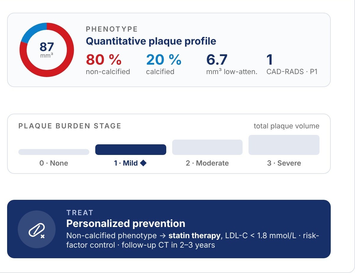
CT shows the disease itself, not only its risk factors. We study how plaque burden and phenotype can guide CT-guided cardiovascular prevention, and how structured, patient-centred reporting can improve treatment initiation and long-term adherence.
Goal: turn every CCTA report into an actionable prevention plan.
CT-based phenotyping and personalized therapy
Drug trials
Longitudinal treatment response
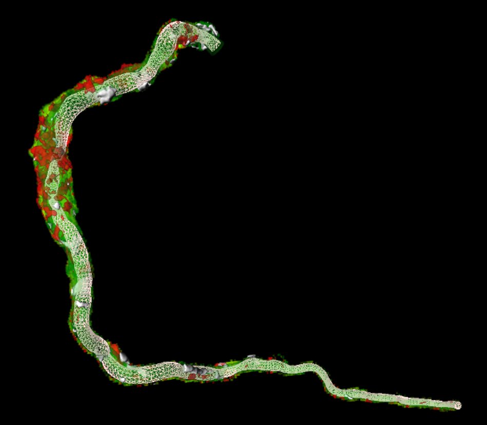
Serial CCTA allows direct measurement of plaque progression and regression under lipid-lowering and anti-inflammatory therapies. Cardio Lab contributes standardised longitudinal plaque analysis to randomised and investigator-initiated trials.
Endpoints include change in total and non-calcified plaque volume, low-attenuation plaque, high-risk features and pericoronary inflammation.
Photon-counting CT & clinical translation
“The question is not whether PCD-CT produces better images, but when better imaging changes the diagnostic pathway, treatment decisions and patient care.”
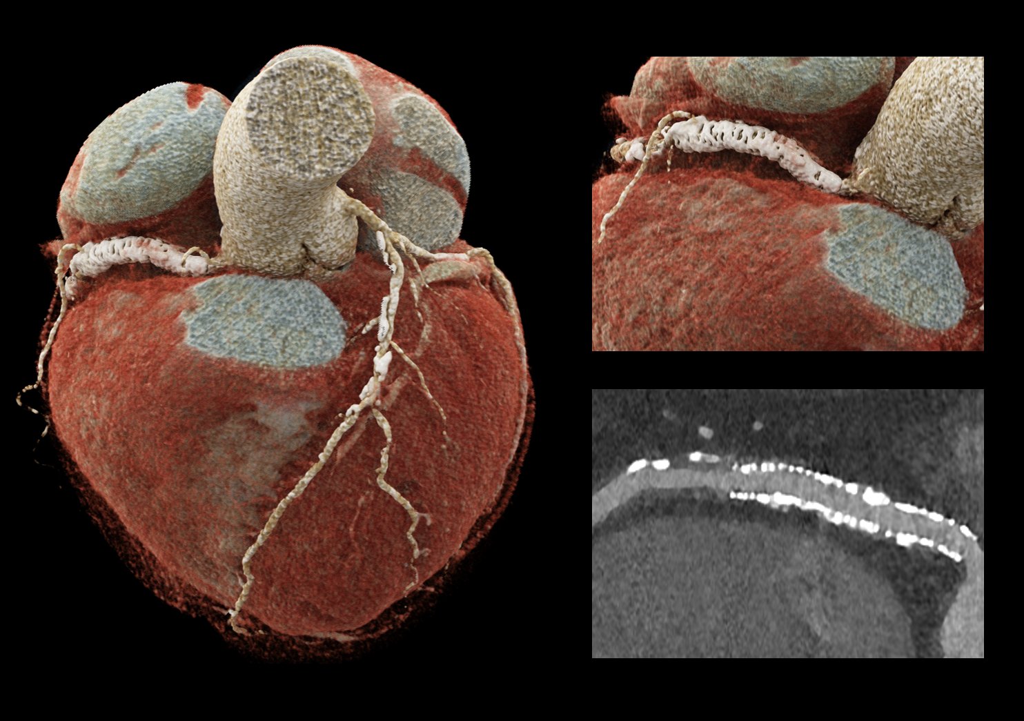
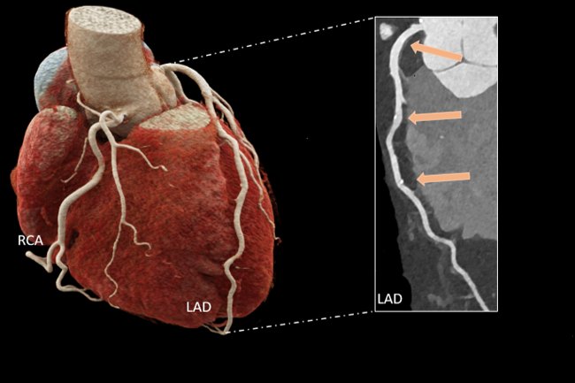
We translate decades of energy-integrating CT evidence to photon-counting CT: ultra-high-resolution coronary and stent imaging, spectral plaque characterisation and reduced blooming — always measured against clinical impact.
Featured: New Eyes for an Old Problem — editorial by B. Szilveszter, JACC: Cardiovascular Imaging, 2026.
Phantom research
Know what you measure
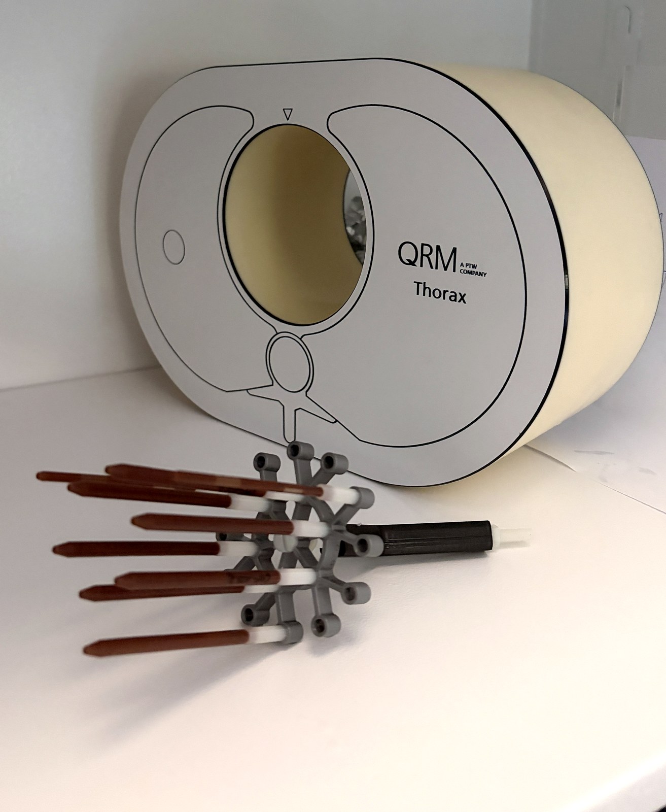
Every quantitative CT biomarker depends on scanner, reconstruction and analysis settings. Using a unique phantom set — in collaboration with George Wesbey — we test plaque and stenosis quantification against known ground truth, so clinical measurements can be trusted.
Ground-truth validation chain
Known ground truth
Acquisition & reconstruction
Quantification
Deviation from truth
Validated protocol
Intervention
CT-guided intervention and procedural planning
CCTA increasingly guides treatment, not only diagnosis. We investigate CT-guided PCI, planning of calcified lesions and stent assessment.
- CT-guided PCI strategy and lesion preparation
- Calcified lesion and stent assessment
- CT-based phenotyping of valvular heart disease
- Aortic disease and post-transplant follow-up
Leadership
Led by clinicians who read the scans
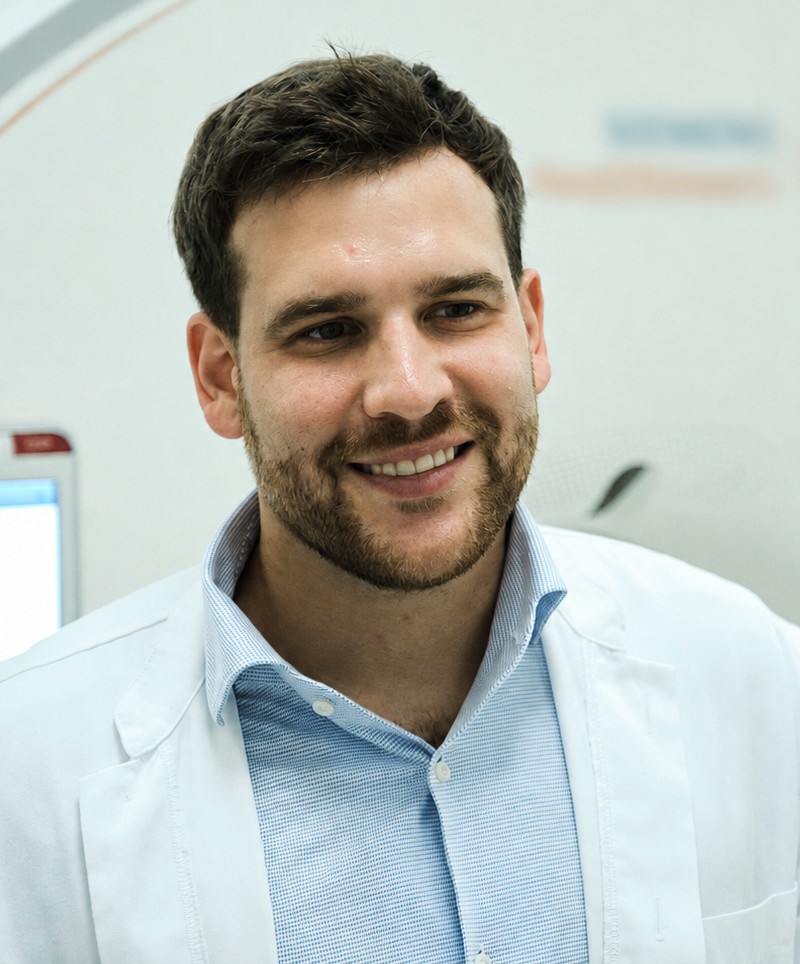
Bálint Szilveszter, MD, PhD, FEACVI, FSCCT is Assistant Professor of Cardiology at the Heart and Vascular Center, Semmelweis University, where he heads the Cardiac CT Working Group and leads the scientific cardiac CT program. He completed his PhD on advanced image reconstruction in coronary CT angiography and has since contributed to international multicentre CT trials, led protocol optimisation for photon-counting CT and mentored numerous young researchers and medical students. His research focuses on CT-guided cardiovascular prevention, quantitative plaque imaging, photon-counting CT and the clinical validation of AI-based plaque analysis tools. He is an EACVI Level III cardiac CT expert and Hungary’s ambassador for the EACVI Heart Imagers of Tomorrow initiative.
Collaborate with Cardio Lab
Core lab services, investigator-initiated studies, AI validation projects or a clinical fellowship — we welcome partners who want CT to change patient care.