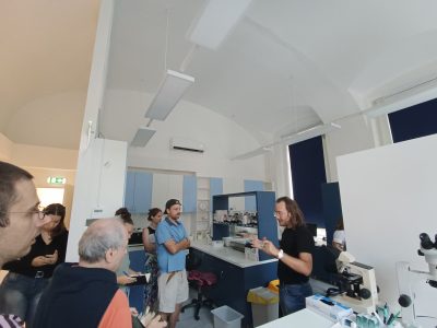
The C80 is one of the ICD (International Classification of Diseases) codes that, when appearing on a histopathology report, can seal a person’s fate. But what does such a code mean? How is it given and why? Who is a histopathology report for, and how is it made? Why does it take even two weeks to get a result from a tumor biopsy when you can have the results of a laboratory test within hours? These were the questions addressed in the first two workshops of the C80 project, led by Richárd Kiss, a pathologist at the Institute of Pathology and Experimental Cancer Research, Semmelweis University, and attended by medical students, cancer patients and clinical oncologists, alongside the visual artists involved in the C80 project.
In the first workshop, participants were given an insight into the biopsy laboratory of the Institute of Pathology and Experimental Cancer Research, where they could follow the journey of a sample from surgery/sampling to the histopathology report. This is a complex process that can require the work of up to a dozen trained medical professionals.
After the surgery/sampling, the sample is delivered to the relevant pathology department/institution by a dedicated health worker and the first step of the workflow, the handover of the material, takes place. At this point, the pathology technician checks the data on the submission form and the specimen container, digitally records the number of specimens, their nature and the contents of the request form. Each sample is given a unique identifier (biopsy number), which allows the sample to be accurately traced. At this point, a pathologist is often needed to decide on the next steps based on the clinical question and the nature of the sample. In most cases, the specimen is received in a fixative called formaldehyde, which preserves the structure of the tissue, thus allowing microscopic examination, but in order for this chemical to penetrate the removed tissue sufficiently, it must be sliced, a task for a medical doctor, as in many cases the outer surface must be marked with dye beforehand. This step is necessary because this marked outer surface is also the so-called resection surface, i.e. if there is intact tissue on this surface, then the abnormal tissue has been removed completely, but if the subsequent microscopic examination shows that the dye has touched the tumour area, the tumour has not been completely removed during the operation.
In formaldehyde, samples usually need to be left for at least 12 hours before processing can continue.
The next step is grossing, during which the formaldehyde-fixed specimen is examined by a pathologist, who describes its appearance (this is also shown on the histological report as a macroscopic description, i.e. what the pathologist sees with his or her eyes without a microscope), takes a photographs and, for larger specimens, decides which parts of the specimen should be sectioned for microscopic examination. The doctor then cuts out a section of the areas he or she considers important, places it in a small plastic cassette and the tissue under examination travels in the plastic cassette.
The tissue samples placed in the cassettes are transferred to a so-called dehydration machine, which immerses the samples in increasingly higher concentrations of alcohol solutions in preparation for the next step, embedding them in paraffin. This process takes several hours.
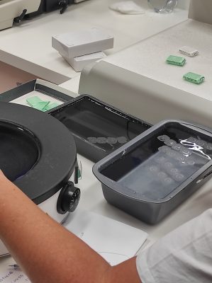
In order to be examined under a light microscope, a tissue sample must be homogeneously solid, which is the purpose of paraffin embedding. Paraffin is a wax-like substance that is liquid when heated but solid at room temperature. In embedding, pathology technicians place the sample in a metal mould, pour liquid paraffin into it and then freeze it on a chilled metal surface. This effectively creates a paraffin cube with the tissue sample contained inside, called a block.
The next step is sectioning. The block is then fixed (clamped) on a cutting machine (microtome) by the technician, who then starts to make the sections using extremely sharp blades. A histological section is 2 to 3 micrometres thick, which is roughly 20 times thinner than a strand of hair. The average human cell is 10-20 micrometres in size, so the histological technician also cuts the cells in half during the sectioning process. A section this thin must be made from each sample so that this almost translucent, membranous section is of uniform thickness and does not tear. This process requires a high degree of precision, manual dexterity, experience and tolerance of monotony, which not all technicians will necessarily be able to do even after specialised training. It is an almost artistic activity, akin to the world of handmade porcelain manufacture.
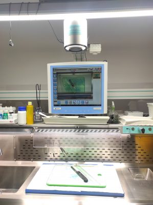
Once a good quality section is made, it is mounted on a glass slide and then painted. Many staining procedures are used in pathological diagnosis, but the most widely used is hematoxylin-eosin staining, which stains the tissue under examination to different shades of pink and blue based on the chemistry (acidic or basic) of the area under examination. After staining, the sections are covered with another glass slide without bubbles, so that the pathologist doctor can obtain them for microscopic examination. The substantive process, where the pathologist determines what disease is visible in the removed tissue, only begins after this lengthy process. Up to this point, at least eight health care workers (specimen supplier, material recipient, evaluation physician, grossing physician, grossing, embedding, sectioning and staining technician) have dealt with the specimen at some level. These are invisible hands whose work is not known from reading a histological report, but without them there is no pathological diagnosis.
How the pathologist decides on the nature of the lesion, what, how and why to record on the histopathology reports after seeing the microscopic image, was the subject of the second workshop of the C80 project in Budapest.
C80 is part of the project “Tackling the taboo of cancer through contemporary art: an integrative education programme that explores the universal human experience of cancer.” funded by the EUniWell (European University of Well Being) Seed Fund. The project is led by Semmelweis University in a consortium with the University of Florence, the University of Murcia and Linnaeus University.
If you find it interesting, share it!